8XC6

Crystal structure of large stokes shift red fluorescent protein tKeima


Experimental Data Snapshot

  • Method: X-RAY DIFFRACTION
  • Resolution: 3.00 Å
  • R-Value Free: 0.247 
  • R-Value Work: 0.196 
  • R-Value Observed: 0.201 

Starting Model: experimental
View more details

wwPDB Validation   3D Report Full Report


This is version 1.1 of the entry. See complete history


Literature

Structural Analysis of the Large Stokes Shift Red Fluorescent Protein tKeima.

Nam, K.H.Xu, Y.

(2024) Molecules 29

  • DOI: https://doi.org/10.3390/molecules29112579
  • Primary Citation of Related Structures:  
    8XC6

  • PubMed Abstract: 

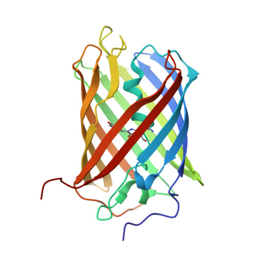
    The Keima family comprises large Stokes shift fluorescent proteins that are useful for dual-color fluorescence cross-correlation spectroscopy and multicolor imaging. The tKeima is a tetrameric large Stokes shift fluorescent protein and serves as the ancestor fluorescent protein for both dKeima and mKeima. The spectroscopic properties of tKeima have been previously reported; however, its structural basis and molecular properties have not yet been elucidated. In this study, we present the crystallographic results of the large Stokes shift fluorescent protein tKeima. The purified tKeima protein spontaneously crystallized after purification without further crystallization. The crystal structure of tKeima was determined at 3.0 Å resolution, revealing a β-barrel fold containing the Gln-Tyr-Gly chromophores mainly with cis-conformation. The tetrameric interfaces of tKeima were stabilized by numerous hydrogen bonds and salt-bridge interactions. These key residues distinguish the substituted residues in dKeima and mKeima. The key structure-based residues involved in the tetramer formation of tKeima provide insights into the generation of a new type of monomeric mKeima. This structural analysis expands our knowledge of the Keima family and provides insights into its protein engineering.


  • Organizational Affiliation

    College of General Education, Kookmin University, Seoul 02707, Republic of Korea.


Macromolecules
Find similar proteins by:  (by identity cutoff)  |  3D Structure
Entity ID: 1
MoleculeChains Sequence LengthOrganismDetailsImage
fluorescent protein
A, B
223Montipora sp. 20Mutation(s): 0 
UniProt
Find proteins for Q1JV72 (Montipora sp. 20)
Explore Q1JV72 
Go to UniProtKB:  Q1JV72
Entity Groups  
Sequence Clusters30% Identity50% Identity70% Identity90% Identity95% Identity100% Identity
UniProt GroupQ1JV72
Sequence Annotations
Expand
  • Reference Sequence
Small Molecules
Modified Residues  1 Unique
IDChains TypeFormula2D DiagramParent
CRQ
Query on CRQ
A, B
L-PEPTIDE LINKINGC16 H16 N4 O5GLN, TYR, GLY
Experimental Data & Validation

Experimental Data

  • Method: X-RAY DIFFRACTION
  • Resolution: 3.00 Å
  • R-Value Free: 0.247 
  • R-Value Work: 0.196 
  • R-Value Observed: 0.201 
  • Space Group: P 2 21 21
Unit Cell:
Length ( Å )Angle ( ˚ )
a = 69.887α = 90
b = 83.504β = 90
c = 109.784γ = 90
Software Package:
Software NamePurpose
REFMACrefinement
HKL-2000data reduction
HKL-2000data scaling
MOLREPphasing

Structure Validation

View Full Validation Report


Entry History & Funding Information

Deposition Data

  • Released Date: 2024-04-17 
  • Deposition Author(s): Nam, K.H.

Funding OrganizationLocationGrant Number
National Research Foundation (NRF, Korea)Korea, Republic OfNRF-2017M3A9F6029736
National Research Foundation (NRF, Korea)Korea, Republic OfNRF-2021R1I1A1A01050838
National Research Foundation (NRF, Korea)Korea, Republic OfNRF-2020M3H1A1075314

Revision History  (Full details and data files)

  • Version 1.0: 2024-04-17
    Type: Initial release
  • Version 1.1: 2024-07-10
    Changes: Database references