


The Crystal Structure of polo-box domain of PLK1 from Biortus
Wang, F., Cheng, W., Yuan, Z., Meng, Q., Zhang, B.To be published.
Experimental Data Snapshot
Starting Model: experimental
View more details
Entity ID: 1 | |||||
|---|---|---|---|---|---|
| Molecule | Chains | Sequence Length | Organism | Details | Image |
| Serine/threonine-protein kinase PLK1 | 237 | Homo sapiens | Mutation(s): 0 Gene Names: PLK1, PLK EC: 2.7.11.21 | 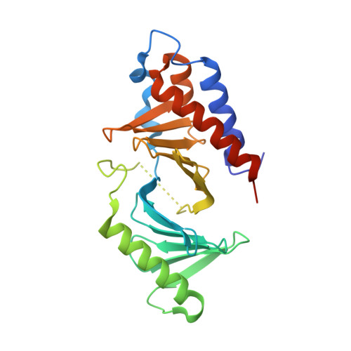 | |
UniProt & NIH Common Fund Data Resources | |||||
PHAROS: P53350 GTEx: ENSG00000166851 | |||||
Entity Groups | |||||
| Sequence Clusters | 30% Identity50% Identity70% Identity90% Identity95% Identity100% Identity | ||||
| UniProt Group | P53350 | ||||
Sequence AnnotationsExpand | |||||
Reference Sequence | |||||
| Ligands 1 Unique | |||||
|---|---|---|---|---|---|
| ID | Chains | Name / Formula / InChI Key | 2D Diagram | 3D Interactions | |
| EDO (Subject of Investigation/LOI) Download:Ideal Coordinates CCD File | B [auth A] | 1,2-ETHANEDIOL C2 H6 O2 LYCAIKOWRPUZTN-UHFFFAOYSA-N |  | ||
| Length ( Å ) | Angle ( ˚ ) |
|---|---|
| a = 33.181 | α = 90 |
| b = 92.786 | β = 101.642 |
| c = 35.89 | γ = 90 |
| Software Name | Purpose |
|---|---|
| REFMAC | refinement |
| XDS | data reduction |
| Aimless | data scaling |
| PHASER | phasing |


