8XAK | pdb_00008xak

Structure of Pif1-G4 complex


Experimental Data Snapshot

  • Method: X-RAY DIFFRACTION
  • Resolution: 3.50 Å
  • R-Value Free: 
    0.324 (Depositor), 0.321 (DCC) 
  • R-Value Work: 
    0.282 (Depositor), 0.283 (DCC) 
  • R-Value Observed: 
    0.286 (Depositor) 

Starting Models: experimental
View more details

wwPDB Validation 3D Report Full Report

Validation slider image for 8XAK

Ligand Structure Quality Assessment 


This is version 1.0 of the entry. See complete history

Literature

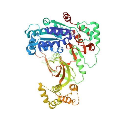
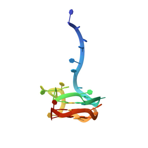
Eukaryotic Pif1 helicase unwinds G-quadruplex and dsDNA using a conserved wedge.

Hong, Z.Byrd, A.K.Gao, J.Das, P.Tan, V.Q.Malone, E.G.Osei, B.Marecki, J.C.Protacio, R.U.Wahls, W.P.Raney, K.D.Song, H.

(2024) Nat Commun 15: 6104-6104

  • DOI: https://doi.org/10.1038/s41467-024-50575-8
  • Primary Citation Related Structures: 
    8XAK

  • PubMed Abstract: 

    G-quadruplexes (G4s) formed by guanine-rich nucleic acids induce genome instability through impeding DNA replication fork progression. G4s are stable DNA structures, the unfolding of which require the functions of DNA helicases. Pif1 helicase binds preferentially to G4 DNA and plays multiple roles in maintaining genome stability, but the mechanism by which Pif1 unfolds G4s is poorly understood. Here we report the co-crystal structure of Saccharomyces cerevisiae Pif1 (ScPif1) bound to a G4 DNA with a 5' single-stranded DNA (ssDNA) segment. Unlike the Thermus oshimai Pif1-G4 structure, in which the 1B and 2B domains confer G4 recognition, ScPif1 recognizes G4 mainly through the wedge region in the 1A domain that contacts the 5' most G-tetrad directly. A conserved Arg residue in the wedge is required for Okazaki fragment processing but not for mitochondrial function or for suppression of gross chromosomal rearrangements. Multiple substitutions at this position have similar effects on resolution of DNA duplexes and G4s, suggesting that ScPif1 may use the same wedge to unwind G4 and dsDNA. Our results reveal the mechanism governing dsDNA unwinding and G4 unfolding by ScPif1 helicase that can potentially be generalized to other eukaryotic Pif1 helicases and beyond.


  • Organizational Affiliation
    • Institute of Molecular and Cell Biology (IMCB), Agency for Science, Technology and Research (A*STAR), Proteos, Singapore, Republic of Singapore.

Macromolecule Content 

  • Total Structure Weight: 146.12 kDa 
  • Atom Count: 9,372 
  • Modeled Residue Count: 1,058 
  • Deposited Residue Count: 1,158 
  • Unique protein chains: 1
  • Unique nucleic acid chains: 1

Macromolecules


Find similar proteins by:|  3D Structure
Entity ID: 1
MoleculeChains  Sequence LengthOrganismDetailsImage
ATP-dependent DNA helicase PIF1A,
B [auth C]
545Saccharomyces cerevisiaeMutation(s): 0 
Gene Names: PIF1
EC: 5.6.2.3
UniProt
Find proteins for P07271 (Saccharomyces cerevisiae (strain ATCC 204508 / S288c))
Explore P07271 
Go to UniProtKB:  P07271
Entity Groups
Sequence Clusters30% Identity50% Identity70% Identity90% Identity95% Identity100% Identity
UniProt GroupP07271
Sequence Annotations
Expand
Reference Sequence
Find similar nucleic acids by:  (by identity cutoff) 
Entity ID: 2
MoleculeChains LengthOrganismImage
DNA (34-MER)C [auth D],
D [auth B]
34synthetic construct
Sequence Annotations
Expand
Reference Sequence

Small Molecules

Ligands 4 Unique
IDChains Name / Formula / InChI Key2D Diagram3D Interactions
ADP
(Subject of Investigation/LOI)

Query on ADP


Download:Ideal Coordinates CCD File
E [auth A],
H [auth C]
ADENOSINE-5'-DIPHOSPHATE
C10 H15 N5 O10 P2
XTWYTFMLZFPYCI-KQYNXXCUSA-N
ALF
(Subject of Investigation/LOI)

Query on ALF


Download:Ideal Coordinates CCD File
F [auth A],
I [auth C]
TETRAFLUOROALUMINATE ION
Al F4
UYOMQIYKOOHAMK-UHFFFAOYSA-J
K
(Subject of Investigation/LOI)

Query on K


Download:Ideal Coordinates CCD File
K [auth D]
L [auth D]
M [auth D]
N [auth B]
O [auth B]
K [auth D],
L [auth D],
M [auth D],
N [auth B],
O [auth B],
P [auth B]
POTASSIUM ION
K
NPYPAHLBTDXSSS-UHFFFAOYSA-N
MG
(Subject of Investigation/LOI)

Query on MG


Download:Ideal Coordinates CCD File
G [auth A],
J [auth C]
MAGNESIUM ION
Mg
JLVVSXFLKOJNIY-UHFFFAOYSA-N

Experimental Data & Validation

Experimental Data

  • Method: X-RAY DIFFRACTION
  • Resolution: 3.50 Å
  • R-Value Free:  0.324 (Depositor), 0.321 (DCC) 
  • R-Value Work:  0.282 (Depositor), 0.283 (DCC) 
  • R-Value Observed: 0.286 (Depositor) 
Space Group: C 1 2 1
Unit Cell:
Length ( Å )Angle ( ˚ )
a = 56.163α = 90
b = 125.141β = 91.47
c = 258.233γ = 90
Software Package:
Software NamePurpose
PHENIXrefinement
XDSdata reduction
XSCALEdata scaling
PHASERphasing

Structure Validation

View Full Validation Report


Ligand Structure Quality Assessment 


Entry History 

& Funding Information

Deposition Data


Funding OrganizationLocationGrant Number
Other governmentSingapore--

Revision History  (Full details and data files)

  • Version 1.0: 2024-07-31
    Type: Initial release