8X41

Crystal structure of DIMT1 in complex with 5'-methylthioadenosine and adenosine from Pyrococcus horikoshii


Experimental Data Snapshot

  • Method: X-RAY DIFFRACTION
  • Resolution: 2.35 Å
  • R-Value Free: 0.205 
  • R-Value Work: 0.165 
  • R-Value Observed: 0.167 

Starting Model: experimental
View more details

wwPDB Validation   3D Report Full Report


Ligand Structure Quality Assessment 


This is version 1.0 of the entry. See complete history


Literature

Structural and functional characterization of archaeal DIMT1 unveils distinct protein dynamics essential for efficient catalysis.

Saha, S.Kanaujia, S.P.

(2024) Structure 

  • DOI: https://doi.org/10.1016/j.str.2024.07.013
  • Primary Citation of Related Structures:  
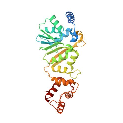
    8X3W, 8X41, 8X44, 8X45, 8X46, 8X47, 8X4G, 8X4I, 8X4L, 8X4O, 8X4P

  • PubMed Abstract: 

    Dimethyladenosine transferase 1 (DIMT1), an ortholog of bacterial KsgA is a conserved protein that assists in ribosome biogenesis by modifying two successive adenosine bases near the 3' end of small subunit (SSU) rRNA. Although KsgA/DIMT1 proteins have been characterized in bacteria and eukaryotes, they are yet unexplored in archaea. Also, their dynamics are not well understood. Here, we structurally and functionally characterized the apo and holo forms of archaeal DIMT1 from Pyrococcus horikoshii. Wild-type protein and mutants were analyzed to capture different transition states, including open, closed, and intermediate states. This study reports a unique inter-domain movement that is needed for substrate (RNA) positioning in the catalytic pocket, and is only observed in the presence of the cognate cofactors S-adenosyl-L-methionine (SAM) or S-adenosyl-L-homocysteine (SAH). The binding of the inhibitor sinefungine, an analog of SAM or SAH, to archaeal DIMT1 blocks the catalytic pocket and renders the enzyme inactive.


  • Organizational Affiliation

    Department of Biosciences and Bioengineering, Indian Institute of Technology Guwahati, Guwahati 781039, Assam, India.


Macromolecules
Find similar proteins by:  (by identity cutoff)  |  3D Structure
Entity ID: 1
MoleculeChains Sequence LengthOrganismDetailsImage
Probable ribosomal RNA small subunit methyltransferase A
A, B
290Pyrococcus horikoshii OT3Mutation(s): 0 
Gene Names: DIMT1
EC: 2.1.1.182
UniProt
Find proteins for O59487 (Pyrococcus horikoshii (strain ATCC 700860 / DSM 12428 / JCM 9974 / NBRC 100139 / OT-3))
Explore O59487 
Go to UniProtKB:  O59487
Entity Groups  
Sequence Clusters30% Identity50% Identity70% Identity90% Identity95% Identity100% Identity
UniProt GroupO59487
Sequence Annotations
Expand
  • Reference Sequence
Small Molecules
Ligands 10 Unique
IDChains Name / Formula / InChI Key2D Diagram3D Interactions
MTA (Subject of Investigation/LOI)
Query on MTA

Download Ideal Coordinates CCD File 
C [auth A],
CA [auth B]
5'-DEOXY-5'-METHYLTHIOADENOSINE
C11 H15 N5 O3 S
WUUGFSXJNOTRMR-IOSLPCCCSA-N
ADN (Subject of Investigation/LOI)
Query on ADN

Download Ideal Coordinates CCD File 
D [auth A],
DA [auth B]
ADENOSINE
C10 H13 N5 O4
OIRDTQYFTABQOQ-KQYNXXCUSA-N
ARG
Query on ARG

Download Ideal Coordinates CCD File 
BA [auth A]ARGININE
C6 H15 N4 O2
ODKSFYDXXFIFQN-BYPYZUCNSA-O
PEG
Query on PEG

Download Ideal Coordinates CCD File 
OA [auth B]DI(HYDROXYETHYL)ETHER
C4 H10 O3
MTHSVFCYNBDYFN-UHFFFAOYSA-N
GOL
Query on GOL

Download Ideal Coordinates CCD File 
KA [auth B]
LA [auth B]
MA [auth B]
NA [auth B]
Q [auth A]
KA [auth B],
LA [auth B],
MA [auth B],
NA [auth B],
Q [auth A],
R [auth A],
S [auth A],
T [auth A],
U [auth A],
V [auth A]
GLYCEROL
C3 H8 O3
PEDCQBHIVMGVHV-UHFFFAOYSA-N
SO3
Query on SO3

Download Ideal Coordinates CCD File 
IA [auth B],
JA [auth B],
O [auth A],
P [auth A]
SULFITE ION
O3 S
LSNNMFCWUKXFEE-UHFFFAOYSA-L
ZN
Query on ZN

Download Ideal Coordinates CCD File 
E [auth A]
EA [auth B]
F [auth A]
FA [auth B]
G [auth A]
E [auth A],
EA [auth B],
F [auth A],
FA [auth B],
G [auth A],
GA [auth B],
H [auth A],
I [auth A],
J [auth A],
K [auth A]
ZINC ION
Zn
PTFCDOFLOPIGGS-UHFFFAOYSA-N
EDO
Query on EDO

Download Ideal Coordinates CCD File 
W [auth A],
X [auth A],
Y [auth A],
Z [auth A]
1,2-ETHANEDIOL
C2 H6 O2
LYCAIKOWRPUZTN-UHFFFAOYSA-N
ACT
Query on ACT

Download Ideal Coordinates CCD File 
AA [auth A],
PA [auth B]
ACETATE ION
C2 H3 O2
QTBSBXVTEAMEQO-UHFFFAOYSA-M
CL
Query on CL

Download Ideal Coordinates CCD File 
HA [auth B],
L [auth A],
M [auth A],
N [auth A]
CHLORIDE ION
Cl
VEXZGXHMUGYJMC-UHFFFAOYSA-M
Experimental Data & Validation

Experimental Data

  • Method: X-RAY DIFFRACTION
  • Resolution: 2.35 Å
  • R-Value Free: 0.205 
  • R-Value Work: 0.165 
  • R-Value Observed: 0.167 
  • Space Group: C 1 2 1
Unit Cell:
Length ( Å )Angle ( ˚ )
a = 120.38α = 90
b = 77.26β = 119.95
c = 94.229γ = 90
Software Package:
Software NamePurpose
REFMACrefinement
HKL-3000data collection
MOSFLMdata reduction
Aimlessdata scaling
PHASERphasing
Cootmodel building
PDB_EXTRACTdata extraction

Structure Validation

View Full Validation Report


Ligand Structure Quality Assessment 


Entry History & Funding Information

Deposition Data


Funding OrganizationLocationGrant Number
Science and Engineering Research Board (SERB)IndiaEEQ/2021/000060

Revision History  (Full details and data files)

  • Version 1.0: 2024-08-28
    Type: Initial release