






 Download Mendeley
The Crystal Structure of PKCi from Biortus
Wang, F., Cheng, W., Lv, Z., Ju, C., Ni, C.To be published.
Experimental Data Snapshot
Starting Model: experimental
View more details
wwPDB Validation 3D Report Full Report

Entity ID: 1 | |||||
|---|---|---|---|---|---|
| Molecule | Chains | Sequence Length | Organism | Details | Image |
| Adenosine 5'-monophosphoramidase HINT1 | 126 | Homo sapiens | Mutation(s): 0 Gene Names: HINT1, HINT, PKCI1, PRKCNH1 EC: 3.9.1 (PDB Primary Data), 3.4.22 (PDB Primary Data) | 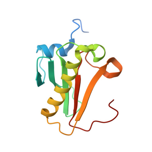 | |
UniProt & NIH Common Fund Data Resources | |||||
PHAROS: P49773 GTEx: ENSG00000169567 | |||||
Entity Groups | |||||
| Sequence Clusters | 30% Identity50% Identity70% Identity90% Identity95% Identity100% Identity | ||||
| UniProt Group | P49773 | ||||
Sequence AnnotationsExpand | |||||
Reference Sequence | |||||
| Length ( Å ) | Angle ( ˚ ) |
|---|---|
| a = 45.76 | α = 90 |
| b = 75.047 | β = 90 |
| c = 80.266 | γ = 90 |
| Software Name | Purpose |
|---|---|
| REFMAC | refinement |
| XDS | data reduction |
| Aimless | data scaling |
| PHASER | phasing |








