8WUT

SpCas9-MMLV RT-pegRNA-target DNA complex (initiation)


Experimental Data Snapshot

  • Method: ELECTRON MICROSCOPY
  • Resolution: 3.00 Å
  • Aggregation State: PARTICLE 
  • Reconstruction Method: SINGLE PARTICLE 

wwPDB Validation   3D Report Full Report


This is version 1.1 of the entry. See complete history


Literature

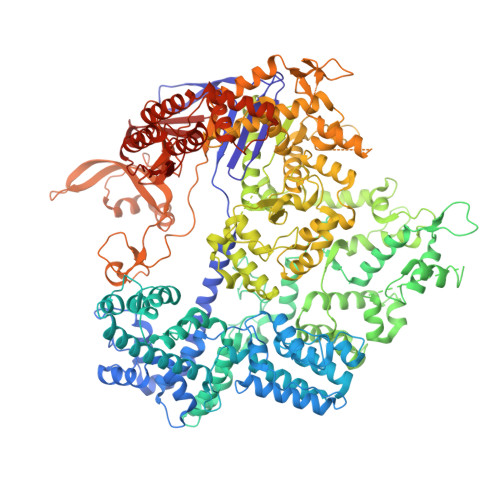
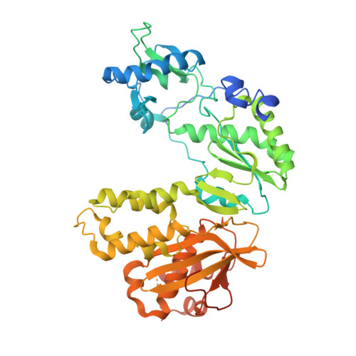
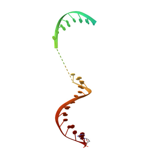
Structural basis for pegRNA-guided reverse transcription by a prime editor.

Shuto, Y.Nakagawa, R.Zhu, S.Hoki, M.Omura, S.N.Hirano, H.Itoh, Y.Zhang, F.Nureki, O.

(2024) Nature 

  • DOI: https://doi.org/10.1038/s41586-024-07497-8
  • Primary Citation of Related Structures:  
    8WUS, 8WUT, 8WUU, 8WUV, 8YGJ

  • PubMed Abstract: 

    The prime editor system composed of Streptococcus pyogenes Cas9 nickase (nSpCas9) and engineered Moloney murine leukaemia virus reverse transcriptase (M-MLV RT) collaborates with a prime editing guide RNA (pegRNA) to facilitate a wide variety of precise genome edits in living cells 1 . However, owing to a lack of structural information, the molecular mechanism of pegRNA-guided reverse transcription by the prime editor remains poorly understood. Here we present cryo-electron microscopy structures of the SpCas9-M-MLV RTΔRNaseH-pegRNA-target DNA complex in multiple states. The termination structure, along with our functional analysis, reveals that M-MLV RT extends reverse transcription beyond the expected site, resulting in scaffold-derived incorporations that cause undesired edits at the target loci. Furthermore, structural comparisons among the pre-initiation, initiation and elongation states show that M-MLV RT remains in a consistent position relative to SpCas9 during reverse transcription, whereas the pegRNA-synthesized DNA heteroduplex builds up along the surface of SpCas9. On the basis of our structural insights, we rationally engineered pegRNA variants and prime-editor variants in which M-MLV RT is fused within SpCas9. Collectively, our findings provide structural insights into the stepwise mechanism of prime editing, and will pave the way for the development of a versatile prime editing toolbox.


  • Organizational Affiliation

    Department of Biological Sciences, Graduate School of Science, The University of Tokyo, Tokyo, Japan.


Macromolecules

Find similar proteins by:  (by identity cutoff)  |  3D Structure
Entity ID: 1
MoleculeChains Sequence LengthOrganismDetailsImage
CRISPR-associated endonuclease Cas9/Csn11,367Streptococcus pyogenes serotype M1Mutation(s): 2 
Gene Names: cas9
UniProt
Find proteins for Q99ZW2 (Streptococcus pyogenes serotype M1)
Explore Q99ZW2 
Go to UniProtKB:  Q99ZW2
Entity Groups  
Sequence Clusters30% Identity50% Identity70% Identity90% Identity95% Identity100% Identity
UniProt GroupQ99ZW2
Sequence Annotations
Expand
  • Reference Sequence
Find similar proteins by:  (by identity cutoff)  |  3D Structure
Entity ID: 2
MoleculeChains Sequence LengthOrganismDetailsImage
Gag-Pol polyproteinB [auth E]496Moloney murine leukemia virusMutation(s): 6 
Gene Names: gag-pro-pol
UniProt
Find proteins for Q8UN00 (Moloney murine leukemia virus)
Explore Q8UN00 
Go to UniProtKB:  Q8UN00
Entity Groups  
Sequence Clusters30% Identity50% Identity70% Identity90% Identity95% Identity100% Identity
UniProt GroupQ8UN00
Sequence Annotations
Expand
  • Reference Sequence
Find similar nucleic acids by: 3D Structure
Entity ID: 3
MoleculeChains LengthOrganismImage
DNA (51-MER)51Streptococcus pyogenes
Sequence Annotations
Expand
  • Reference Sequence

Find similar nucleic acids by:  Sequence   |   3D Structure  

Entity ID: 4
MoleculeChains LengthOrganismImage
DNA (5'-D(*TP*GP*AP*TP*GP*GP*CP*AP*GP*AP*GP*TP*AP*CP*TP*AP*G)-3')17Streptococcus pyogenes
Sequence Annotations
Expand
  • Reference Sequence
Find similar nucleic acids by: 3D Structure
Entity ID: 5
MoleculeChains LengthOrganismImage
RNA (115-MER)E [auth B]115Streptococcus pyogenes
Sequence Annotations
Expand
  • Reference Sequence
Find similar nucleic acids by: 3D Structure
Entity ID: 6
MoleculeChains LengthOrganismImage
DNA/RNA (35-MER)35Streptococcus pyogenes
Sequence Annotations
Expand
  • Reference Sequence
Experimental Data & Validation

Experimental Data

  • Method: ELECTRON MICROSCOPY
  • Resolution: 3.00 Å
  • Aggregation State: PARTICLE 
  • Reconstruction Method: SINGLE PARTICLE 

Structure Validation

View Full Validation Report


Entry History & Funding Information

Deposition Data


Funding OrganizationLocationGrant Number
Japan Agency for Medical Research and Development (AMED)JapanJP23ama121012

Revision History  (Full details and data files)

  • Version 1.0: 2024-06-05
    Type: Initial release
  • Version 1.1: 2024-06-12
    Changes: Data collection, Database references, Structure summary