Structural determinants of spike infectivity in bat SARS-like coronaviruses RsSHC014 and WIV1.
Qiao, S., Wang, X.(2024) J Virol 98: e0034224-e0034224
- PubMed: 39028202 Search on PubMed
- DOI: https://doi.org/10.1128/jvi.00342-24
- Primary Citation Related Structures:
8WLU, 8WLY, 8WLZ, 8WQ0 - PubMed Abstract:
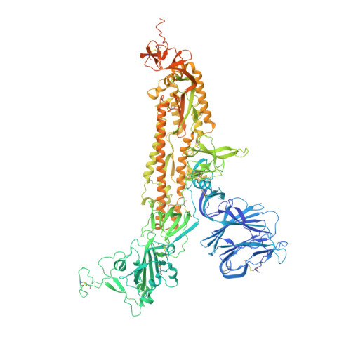
The recurrent spillovers of coronaviruses (CoVs) have posed severe threats to public health and the global economy. Bat severe acute respiratory syndrome (SARS)-like CoVs RsSHC014 and WIV1, currently circulating in bat populations, are poised for human emergence. The trimeric spike (S) glycoprotein, responsible for receptor recognition and membrane fusion, plays a critical role in cross-species transmission and infection. Here, we determined the cryo-electron microscopy (EM) structures of the RsSHC014 S protein in the closed state at 2.9 Å, the WIV1 S protein in the closed state at 2.8 Å, and the intermediate state at 4.0 Å. In the intermediate state, one receptor-binding domain (RBD) is in the "down" position, while the other two RBDs exhibit poor density. We also resolved the complex structure of the WIV1 S protein bound to human ACE2 (hACE2) at 4.5 Å, which provides structural basis for the future emergence of WIV1 in humans. Through biochemical experiments, we found that despite strong binding affinities between the RBDs and both human and civet ACE2, the pseudoviruses of RsSHC014, but not WIV1, failed to infect 293T cells overexpressing either human or civet ACE2. Mutagenesis analysis revealed that the Y623H substitution, located in the SD2 region, significantly improved the cell entry efficiency of RsSHC014 pseudoviruses, which is likely accomplished by promoting the open conformation of spike glycoproteins. Our findings emphasize the necessity of both efficient RBD lifting and tight RBD-hACE2 binding for viral infection and underscore the significance of the 623 site of the spike glycoprotein for the infectivity of bat SARS-like CoVs. The bat SARS-like CoVs RsSHC014 and WIV1 can use hACE2 for cell entry without further adaptation, indicating their potential risk of emergence in human populations. The S glycoprotein, responsible for receptor recognition and membrane fusion, plays a crucial role in cross-species transmission and infection. In this study, we determined the cryo-EM structures of the S glycoproteins of RsSHC014 and WIV1. Detailed comparisons revealed dynamic structural variations within spike proteins. We also elucidated the complex structure of WIV1 S-hACE2, providing structural evidence for the potential emergence of WIV1 in humans. Although RsSHC014 and WIV1 had similar hACE2-binding affinities, they exhibited distinct pseudovirus cell entry behavior. Through mutagenesis and cryo-EM analysis, we revealed that besides the structural variations, the 623 site in the SD2 region is another important structural determinant of spike infectivity.
- The Ministry of Education Key Laboratory of Protein Science, Beijing, China.
Organizational Affiliation: