


Cryo-EM structure of a bacterial protein
Yu, G., Liao, F., Li, X., Li, Z., Zhang, H.To be published.
Experimental Data Snapshot
wwPDB Validation 3D Report Full Report

Entity ID: 1 | |||||
|---|---|---|---|---|---|
| Molecule | Chains | Sequence Length | Organism | Details | Image |
| Helicase HerA central domain-containing protein | A [auth D], B [auth M], C [auth N], D [auth O], E [auth L] | 696 | Paenibacillus sp. 453mf | Mutation(s): 0 Gene Names: SAMN04488601_104156 | 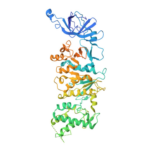 |
| Ligands 2 Unique | |||||
|---|---|---|---|---|---|
| ID | Chains | Name / Formula / InChI Key | 2D Diagram | 3D Interactions | |
| AGS (Subject of Investigation/LOI) Download:Ideal Coordinates CCD File | F [auth M], H [auth O], J [auth L] | PHOSPHOTHIOPHOSPHORIC ACID-ADENYLATE ESTER C10 H16 N5 O12 P3 S NLTUCYMLOPLUHL-KQYNXXCUSA-N |  | ||
| MG (Subject of Investigation/LOI) Download:Ideal Coordinates CCD File | G [auth M], I [auth O], K [auth L] | MAGNESIUM ION Mg JLVVSXFLKOJNIY-UHFFFAOYSA-N |  | ||
| Task | Software Package | Version |
|---|---|---|
| MODEL REFINEMENT | PHENIX | |
| Funding Organization | Location | Grant Number |
|---|---|---|
| National Natural Science Foundation of China (NSFC) | China | 32200496 |


