8WHI

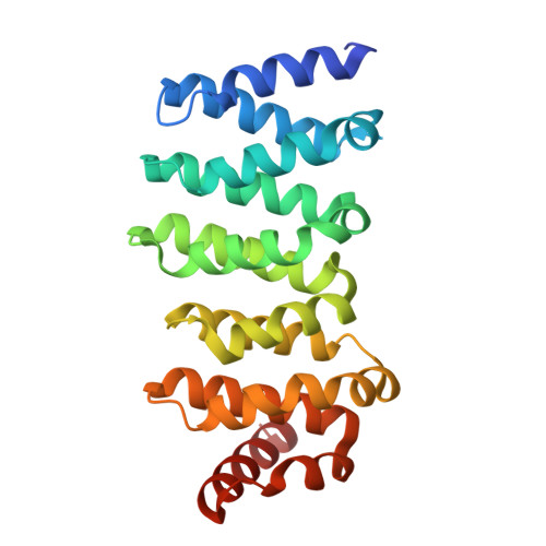
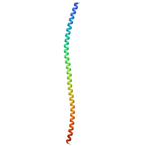
Crystal structure of native CLASP2 in complex with CLIP170


Experimental Data Snapshot

  • Method: X-RAY DIFFRACTION
  • Resolution: 1.85 Å
  • R-Value Free: 0.237 
  • R-Value Work: 0.191 
  • R-Value Observed: 0.193 

Starting Model: experimental
View more details

wwPDB Validation   3D Report Full Report


This is version 1.0 of the entry. See complete history


Literature

CLASP-mediated competitive binding in protein condensates directs microtubule growth.

Jia, X.Lin, L.Guo, S.Zhou, L.Jin, G.Dong, J.Xiao, J.Xie, X.Li, Y.He, S.Wei, Z.Yu, C.

(2024) Nat Commun 15: 6509-6509

  • DOI: https://doi.org/10.1038/s41467-024-50863-3
  • Primary Citation of Related Structures:  
    8WHH, 8WHI, 8WHJ, 8WHK, 8WHL, 8WHM

  • PubMed Abstract: 

    Microtubule organization in cells relies on targeting mechanisms. Cytoplasmic linker proteins (CLIPs) and CLIP-associated proteins (CLASPs) are key regulators of microtubule organization, yet the underlying mechanisms remain elusive. Here, we reveal that the C-terminal domain of CLASP2 interacts with a common motif found in several CLASP-binding proteins. This interaction drives the dynamic localization of CLASP2 to distinct cellular compartments, where CLASP2 accumulates in protein condensates at the cell cortex or the microtubule plus end. These condensates physically contact each other via CLASP2-mediated competitive binding, determining cortical microtubule targeting. The phosphorylation of CLASP2 modulates the dynamics of the condensate-condensate interaction and spatiotemporally navigates microtubule growth. Moreover, we identify additional CLASP-interacting proteins that are involved in condensate contacts in a CLASP2-dependent manner, uncovering a general mechanism governing microtubule targeting. Our findings not only unveil a tunable multiphase system regulating microtubule organization, but also offer general mechanistic insights into intricate protein-protein interactions at the mesoscale level.


  • Organizational Affiliation

    Shenzhen Key Laboratory of Biomolecular Assembling and Regulation, Shenzhen, Guangdong, 518055, China.


Macromolecules
Find similar proteins by:  (by identity cutoff)  |  3D Structure
Entity ID: 1
MoleculeChains Sequence LengthOrganismDetailsImage
CLIP-associating protein 2235Homo sapiensMutation(s): 0 
Gene Names: CLASP2KIAA0627
UniProt & NIH Common Fund Data Resources
Find proteins for O75122 (Homo sapiens)
Explore O75122 
Go to UniProtKB:  O75122
PHAROS:  O75122
GTEx:  ENSG00000163539 
Entity Groups  
Sequence Clusters30% Identity50% Identity70% Identity90% Identity95% Identity100% Identity
UniProt GroupO75122
Sequence Annotations
Expand
  • Reference Sequence
Find similar proteins by:  (by identity cutoff)  |  3D Structure
Entity ID: 2
MoleculeChains Sequence LengthOrganismDetailsImage
CLIP1 proteinB [auth E],
C [auth F]
111Homo sapiensMutation(s): 0 
Gene Names: CLIP1
UniProt & NIH Common Fund Data Resources
Find proteins for P30622 (Homo sapiens)
Explore P30622 
Go to UniProtKB:  P30622
PHAROS:  P30622
GTEx:  ENSG00000130779 
Entity Groups  
Sequence Clusters30% Identity50% Identity70% Identity90% Identity95% Identity100% Identity
UniProt GroupP30622
Sequence Annotations
Expand
  • Reference Sequence
Experimental Data & Validation

Experimental Data

  • Method: X-RAY DIFFRACTION
  • Resolution: 1.85 Å
  • R-Value Free: 0.237 
  • R-Value Work: 0.191 
  • R-Value Observed: 0.193 
  • Space Group: P 21 21 21
Unit Cell:
Length ( Å )Angle ( ˚ )
a = 52.624α = 90
b = 85.545β = 90
c = 114.195γ = 90
Software Package:
Software NamePurpose
PHENIXrefinement
HKL-2000data scaling
HKL-2000data reduction
PHASERphasing

Structure Validation

View Full Validation Report


Entry History & Funding Information

Deposition Data


Funding OrganizationLocationGrant Number
National Natural Science Foundation of China (NSFC)China31971131
National Natural Science Foundation of China (NSFC)China32170697
National Natural Science Foundation of China (NSFC)China32370743
National Natural Science Foundation of China (NSFC)China32371009

Revision History  (Full details and data files)

  • Version 1.0: 2024-08-28
    Type: Initial release