Protein Allostery Study in Cells Using NMR Spectroscopy.
Chen, X., Zhang, X., Qin, M., Chen, J., Wang, M., Liu, Z., An, L., Song, X., Yao, L.(2024) Anal Chem 96: 7065-7072
- PubMed: 38652079 Search on PubMed
- DOI: https://doi.org/10.1021/acs.analchem.4c00360
- Primary Citation Related Structures:
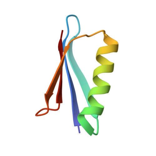
8WCJ - PubMed Abstract:
Protein allostery is commonly observed in vitro. But how protein allostery behaves in cells is unknown. In this work, a protein monomer-dimer equilibrium system was built with the allosteric effect on the binding characterized using NMR spectroscopy through mutations away from the dimer interface. A chemical shift linear fitting method was developed that enabled us to accurately determine the dissociation constant. A total of 28 allosteric mutations were prepared and grouped to negative allosteric, nonallosteric, and positive allosteric modulators. ∼ 50% of mutations displayed the allosteric-state changes when moving from a buffered solution into cells. For example, there were no positive allosteric modulators in the buffered solution but eight in cells. The change in protein allostery is correlated with the interactions between the protein and the cellular environment. These interactions presumably drive the surrounding macromolecules in cells to transiently bind to the monomer and dimer mutational sites and change the free energies of the two species differently which generate new allosteric effects. These surrounding macromolecules create a new protein allostery pathway that is only present in cells.
- Qingdao Institute of Bioenergy and Bioprocess Technology, Chinese Academy of Sciences, Qingdao 266101, China.
Organizational Affiliation: