Three-dimensional architecture of ESCRT-III flat spirals on the membrane.
Liu, M., Liu, Y., Song, T., Yang, L., Qi, L., Zhang, Y.Z., Wang, Y., Shen, Q.T.(2024) Proc Natl Acad Sci U S A 121: e2319115121-e2319115121
- PubMed: 38709931 Search on PubMedSearch on PubMed Central
- DOI: https://doi.org/10.1073/pnas.2319115121
- Primary Citation Related Structures:
8WB6, 8WB7 - PubMed Abstract:
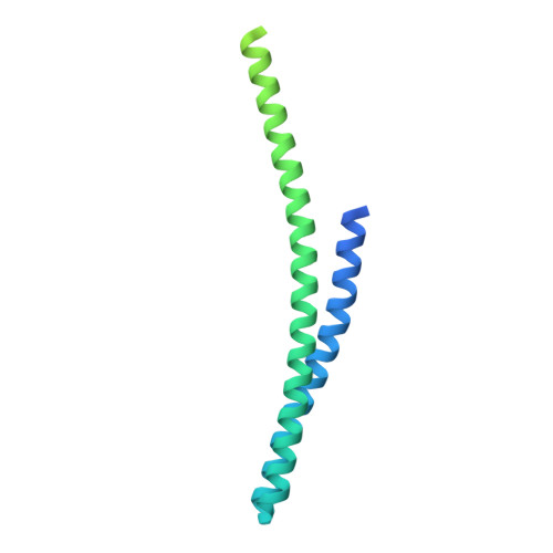
The endosomal sorting complexes required for transport (ESCRTs) are responsible for membrane remodeling in many cellular processes, such as multivesicular body biogenesis, viral budding, and cytokinetic abscission. ESCRT-III, the most abundant ESCRT subunit, assembles into flat spirals as the primed state, essential to initiate membrane invagination. However, the three-dimensional architecture of ESCRT-III flat spirals remained vague for decades due to highly curved filaments with a small diameter and a single preferred orientation on the membrane. Here, we unveiled that yeast Snf7, a component of ESCRT-III, forms flat spirals on the lipid monolayers using cryogenic electron microscopy. We developed a geometry-constrained Euler angle-assigned reconstruction strategy and obtained moderate-resolution structures of Snf7 flat spirals with varying curvatures. Our analyses showed that Snf7 subunits recline on the membrane with N-terminal motifs α0 as anchors, adopt an open state with fused α2/3 helices, and bend α2/3 gradually from the outer to inner parts of flat spirals. In all, we provide the orientation and conformations of ESCRT-III flat spirals on the membrane and unveil the underlying assembly mechanism, which will serve as the initial step in understanding how ESCRTs drive membrane abscission.
- School of Life Sciences, Department of Chemical Biology, Southern University of Science and Technology, Shenzhen 518055, China.
Organizational Affiliation: