


Multifaceted Role of the Substrate Phosphate Group in Transketolase Catalysis
Liu, Z., Xiao, C., Lin, S., Tittmann, K., Dai, S.(2024) ACS Catal 14: 355-365
Experimental Data Snapshot
Starting Model: experimental
View more details
(2024) ACS Catal 14: 355-365
Entity ID: 1 | |||||
|---|---|---|---|---|---|
| Molecule | Chains | Sequence Length | Organism | Details | Image |
| Transketolase | 637 | Homo sapiens | Mutation(s): 0 Gene Names: TKT EC: 2.2.1.1 | 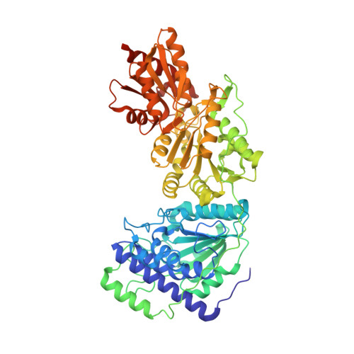 | |
UniProt & NIH Common Fund Data Resources | |||||
PHAROS: P29401 GTEx: ENSG00000163931 | |||||
Entity Groups | |||||
| Sequence Clusters | 30% Identity50% Identity70% Identity90% Identity95% Identity100% Identity | ||||
| UniProt Group | P29401 | ||||
Sequence AnnotationsExpand | |||||
Reference Sequence | |||||
| Ligands 4 Unique | |||||
|---|---|---|---|---|---|
| ID | Chains | Name / Formula / InChI Key | 2D Diagram | 3D Interactions | |
| THD (Subject of Investigation/LOI) Download:Ideal Coordinates CCD File | E [auth A], S [auth B] | 2-[3-[(4-AMINO-2-METHYL-5-PYRIMIDINYL)METHYL]-2-(1,2-DIHYDROXYETHYL)-4-METHYL-1,3-THIAZOL-3-IUM-5-YL]ETHYL TRIHYDROGEN
DIPHOSPHATE C14 H22 N4 O9 P2 S LXZUEFPJZTWGEL-SDNWHVSQSA-N |  | ||
| 3GR (Subject of Investigation/LOI) Download:Ideal Coordinates CCD File | D [auth A], R [auth B] | D-Glyceraldehyde C3 H6 O3 MNQZXJOMYWMBOU-VKHMYHEASA-N |  | ||
| EDO Download:Ideal Coordinates CCD File | AA [auth B] BA [auth B] CA [auth B] DA [auth B] EA [auth B] | 1,2-ETHANEDIOL C2 H6 O2 LYCAIKOWRPUZTN-UHFFFAOYSA-N |  | ||
| CA Download:Ideal Coordinates CCD File | C [auth A], Q [auth B] | CALCIUM ION Ca BHPQYMZQTOCNFJ-UHFFFAOYSA-N |  | ||
| Length ( Å ) | Angle ( ˚ ) |
|---|---|
| a = 73.5 | α = 90 |
| b = 86.55 | β = 93.85 |
| c = 92.7 | γ = 90 |
| Software Name | Purpose |
|---|---|
| XDS | data reduction |
| XSCALE | data scaling |
| PHENIX | phasing |
| PHENIX | refinement |
| Funding Organization | Location | Grant Number |
|---|---|---|
| German Research Foundation (DFG) | Germany | FOR 1296/TP 3 |
| National Natural Science Foundation of China (NSFC) | China | 32271305 |


