8W9X | pdb_00008w9x

Structure of a bacterial protein


Experimental Data Snapshot

  • Method: ELECTRON MICROSCOPY
  • Resolution: 4.40 Å
  • Aggregation State: PARTICLE 
  • Reconstruction Method: SINGLE PARTICLE 

wwPDB Validation 3D Report Full Report

Validation slider image for 8W9X

This is version 1.1 of the entry. See complete history

Literature

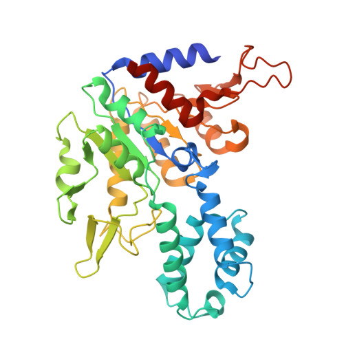
structure of a bacterial protein

Yu, G.Liao, F.Li, X.Li, Z.Zhang, H.

To be published.

Macromolecule Content 

  • Total Structure Weight: 523.31 kDa 
  • Atom Count: 32,616 
  • Modeled Residue Count: 4,320 
  • Deposited Residue Count: 4,572 
  • Unique protein chains: 1

Macromolecules

Find similar proteins by:|  3D Structure
Entity ID: 1
MoleculeChains  Sequence LengthOrganismDetailsImage
SIR2-like domain-containing protein
A, B, C, D, E
A, B, C, D, E, F, G, H, I, J, K, L
381Paenibacillus sp. 453mfMutation(s): 0 
Gene Names: SAMN04488601_104157
UniProt
Find proteins for A0ACD6BAK5 (Paenibacillus sp. 453mf)
Explore A0ACD6BAK5 
Go to UniProtKB:  A0ACD6BAK5
Entity Groups
Sequence Clusters30% Identity50% Identity70% Identity90% Identity95% Identity100% Identity
UniProt GroupA0ACD6BAK5
Sequence Annotations
Expand
Reference Sequence

Experimental Data & Validation

Experimental Data

  • Method: ELECTRON MICROSCOPY
  • Resolution: 4.40 Å
  • Aggregation State: PARTICLE 
  • Reconstruction Method: SINGLE PARTICLE 
EM Software:
TaskSoftware PackageVersion
MODEL REFINEMENTPHENIX

Structure Validation

View Full Validation Report


Entry History 

& Funding Information

Deposition Data


Funding OrganizationLocationGrant Number
National Natural Science Foundation of China (NSFC)China32200496

Revision History  (Full details and data files)

  • Version 1.0: 2024-09-11
    Type: Initial release
  • Version 1.1: 2025-07-02
    Changes: Data collection, Structure summary