8W95 | pdb_00008w95

Azumapecten Farreri homopolymeric ferritin (ApF) mutant-H2KE exposed to H2O2 for 20 s


Experimental Data Snapshot

  • Method: X-RAY DIFFRACTION
  • Resolution: 2.03 Å
  • R-Value Free: 
    0.227 (Depositor), 0.228 (DCC) 
  • R-Value Work: 
    0.186 (Depositor), 0.187 (DCC) 
  • R-Value Observed: 
    0.190 (Depositor) 

Starting Model: experimental
View more details

wwPDB Validation 3D Report Full Report

Validation slider image for 8W95

This is version 1.1 of the entry. See complete history

Literature

Structural Insights into the Reaction between Hydrogen Peroxide and Di-iron Complexes at the Ferroxidase Center of Ferritin.

Jiao, R.Zhao, G.Zhang, T.

(2024) Inorg Chem 63: 3359-3365

  • DOI: https://doi.org/10.1021/acs.inorgchem.3c03889
  • Primary Citation Related Structures: 
    8W91, 8W92, 8W93, 8W94, 8W95, 8WB3

  • PubMed Abstract: 

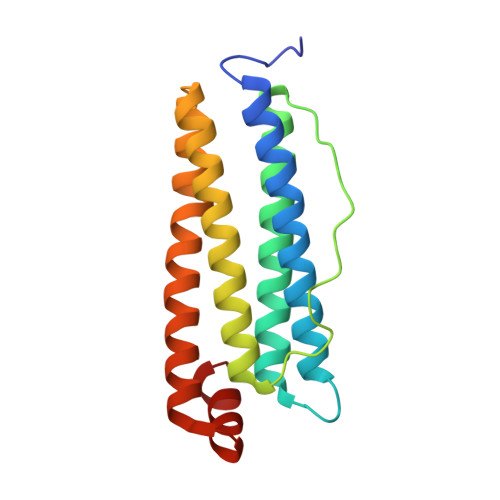
    The Fe(II) oxidation mechanism in the ferroxidase center of heavy chain ferritin has been studied extensively. However, the actual production of H 2 O 2 was found to be substantially lower than expected at low flux of Fe(II) to ferritin subunits. Here, we demonstrated that H 2 O 2 could interact with the di-iron nuclear center, leading to the production of hydroxyl radicals and oxygen. Two reaction intermediates were captured in the ferroxidase center by using the time-lapse crystallographic techniques in a shellfish ferritin. The crystal structures revealed the binding of H 2 O 2 as a μ -1,2-peroxo-diferric species and the binding of O 2 to the diferric structure. This investigation sheds light on the reaction between the di-iron nuclear center and H 2 O 2 and provides insights for the exploitation of metalloenzymes.


  • Organizational Affiliation
    • Beijing Key Laboratory of Functional Food from Plant Resources, College of Food Science and Nutritional Engineering, China Agricultural University, Beijing 100083, China.

Macromolecule Content 

  • Total Structure Weight: 20.16 kDa 
  • Atom Count: 1,519 
  • Modeled Residue Count: 169 
  • Deposited Residue Count: 171 
  • Unique protein chains: 1

Macromolecules

Find similar proteins by:|  3D Structure
Entity ID: 1
MoleculeChains  Sequence LengthOrganismDetailsImage
FerritinA [auth b]171Azumapecten farreriMutation(s): 2 
EC: 1.16.3.1
UniProt
Find proteins for A0A173CSP7 (Azumapecten farreri)
Explore A0A173CSP7 
Go to UniProtKB:  A0A173CSP7
Entity Groups
Sequence Clusters30% Identity50% Identity70% Identity90% Identity95% Identity100% Identity
UniProt GroupA0A173CSP7
Sequence Annotations
Expand
Reference Sequence

Experimental Data & Validation

Experimental Data

  • Method: X-RAY DIFFRACTION
  • Resolution: 2.03 Å
  • R-Value Free:  0.227 (Depositor), 0.228 (DCC) 
  • R-Value Work:  0.186 (Depositor), 0.187 (DCC) 
  • R-Value Observed: 0.190 (Depositor) 
Space Group: I 4 3 2
Unit Cell:
Length ( Å )Angle ( ˚ )
a = 148.641α = 90
b = 148.641β = 90
c = 148.641γ = 90
Software Package:
Software NamePurpose
PHENIXrefinement
XDSdata reduction
XDSdata scaling
MOLREPphasing

Structure Validation

View Full Validation Report


Entry History 

& Funding Information

Deposition Data


Funding OrganizationLocationGrant Number
National Natural Science Foundation of China (NSFC)China--

Revision History  (Full details and data files)

  • Version 1.0: 2024-09-11
    Type: Initial release
  • Version 1.1: 2025-03-26
    Changes: Database references, Structure summary