8W33

Structure of McrD (methyl-coenzyme M reductase operon protein D) from Methanomassiliicoccus luminyensis


Experimental Data Snapshot

  • Method: X-RAY DIFFRACTION
  • Resolution: 1.65 Å
  • R-Value Free: 0.192 
  • R-Value Work: 0.178 

wwPDB Validation   3D Report Full Report


This is version 1.2 of the entry. See complete history


Literature

The crystal structure of methanogen McrD, a methyl-coenzyme M reductase-associated protein.

Sutherland-Smith, A.J.Carbone, V.Schofield, L.R.Cronin, B.Duin, E.C.Ronimus, R.S.

(2024) FEBS Open Bio 14: 1222-1229

  • DOI: https://doi.org/10.1002/2211-5463.13848
  • Primary Citation of Related Structures:  
    8W33

  • PubMed Abstract: 

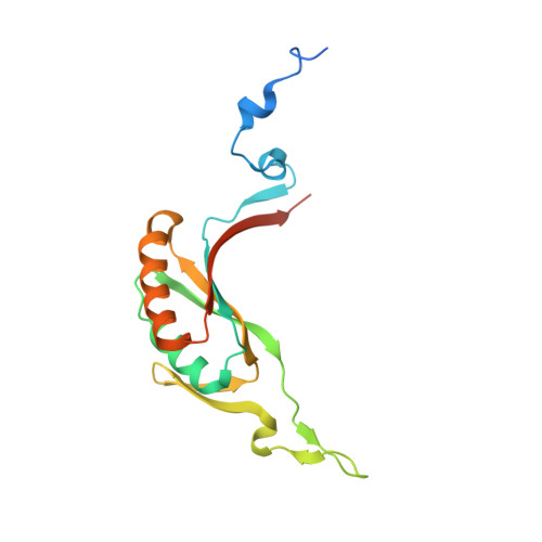
    Methyl-coenzyme M reductase (MCR) is a multi-subunit (α 2 β 2 γ 2 ) enzyme responsible for methane formation via its unique F 430 cofactor. The genes responsible for producing MCR (mcrA, mcrB and mcrG) are typically colocated with two other highly conserved genes mcrC and mcrD. We present here the high-resolution crystal structure for McrD from a human gut methanogen Methanomassiliicoccus luminyensis strain B10. The structure reveals that McrD comprises a ferredoxin-like domain assembled into an α + β barrel-like dimer with conformational flexibility exhibited by a functional loop. The description of the M. luminyensis McrD crystal structure contributes to our understanding of this key conserved methanogen protein typically responsible for promoting MCR activity and the production of methane, a greenhouse gas.


  • Organizational Affiliation

    School of Natural Sciences, Massey University, Palmerston North, New Zealand.


Macromolecules
Find similar proteins by:  (by identity cutoff)  |  3D Structure
Entity ID: 1
MoleculeChains Sequence LengthOrganismDetailsImage
McrD (methyl-coenzyme M reductase operon protein D)
A, B
172Methanomassiliicoccus luminyensis B10Mutation(s): 0 
Gene Names: mcrD
Entity Groups  
Sequence Clusters30% Identity50% Identity70% Identity90% Identity95% Identity100% Identity
Sequence Annotations
Expand
  • Reference Sequence
Experimental Data & Validation

Experimental Data

  • Method: X-RAY DIFFRACTION
  • Resolution: 1.65 Å
  • R-Value Free: 0.192 
  • R-Value Work: 0.178 
  • Space Group: C 2 2 21
Unit Cell:
Length ( Å )Angle ( ˚ )
a = 86.062α = 90
b = 90.224β = 90
c = 121.415γ = 90
Software Package:
Software NamePurpose
REFMACrefinement
Cootmodel building
XDSdata reduction
Aimlessdata scaling
BUCCANEERmodel building
CRANK2phasing

Structure Validation

View Full Validation Report


Entry History & Funding Information

Deposition Data


Funding OrganizationLocationGrant Number
Other governmentNew Zealand--

Revision History  (Full details and data files)

  • Version 1.0: 2024-07-03
    Type: Initial release
  • Version 1.1: 2024-08-21
    Changes: Database references
  • Version 1.2: 2024-08-28
    Changes: Database references