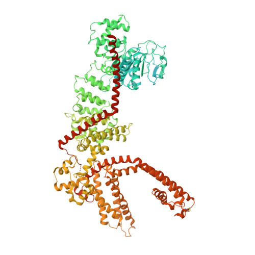
8W2L

TRPM7 structure in complex with anticancer agent CCT128930 in closed state

  • Classification: MEMBRANE PROTEIN
  • Organism(s): Aequorea victoria, Mus musculus
  • Expression System: Homo sapiens
  • Mutation(s): No 
  • Membrane Protein: Yes  PDBTMMemProtMDmpstruc

  • Deposited: 2024-02-20 Released: 2024-04-17 
  • Deposition Author(s): Nadezhdin, K.D., Sobolevsky, A.I.
  • Funding Organization(s): National Institutes of Health/National Institute of Arthritis and Musculoskeletal and Skin Diseases (NIH/NIAMS), National Institutes of Health/National Cancer Institute (NIH/NCI), National Institutes of Health/National Institute of Neurological Disorders and Stroke (NIH/NINDS), German Research Foundation (DFG)

Experimental Data Snapshot

  • Method: ELECTRON MICROSCOPY
  • Resolution: 2.45 Å
  • Aggregation State: PARTICLE 
  • Reconstruction Method: SINGLE PARTICLE 

wwPDB Validation   3D Report Full Report


This is version 1.2 of the entry. See complete history


Literature

Structural basis of selective TRPM7 inhibition by the anticancer agent CCT128930.

Nadezhdin, K.D.Correia, L.Shalygin, A.Aktolun, M.Neuberger, A.Gudermann, T.Kurnikova, M.G.Chubanov, V.Sobolevsky, A.I.

(2024) Cell Rep 43: 114108-114108

  • DOI: https://doi.org/10.1016/j.celrep.2024.114108
  • Primary Citation of Related Structures:  
    8W2L

  • PubMed Abstract: 

    TRP channels are implicated in various diseases, but high structural similarity between them makes selective pharmacological modulation challenging. Here, we study the molecular mechanism underlying specific inhibition of the TRPM7 channel, which is essential for cancer cell proliferation, by the anticancer agent CCT128930 (CCT). Using cryo-EM, functional analysis, and MD simulations, we show that CCT binds to a vanilloid-like (VL) site, stabilizing TRPM7 in the closed non-conducting state. Similar to other allosteric inhibitors of TRPM7, NS8593 and VER155008, binding of CCT is accompanied by displacement of a lipid that resides in the VL site in the apo condition. Moreover, we demonstrate the principal role of several residues in the VL site enabling CCT to inhibit TRPM7 without impacting the homologous TRPM6 channel. Hence, our results uncover the central role of the VL site for the selective interaction of TRPM7 with small molecules that can be explored in future drug design.


  • Organizational Affiliation

    Department of Biochemistry and Molecular Biophysics, Columbia University, New York, NY, USA.


Macromolecules
Find similar proteins by:  (by identity cutoff)  |  3D Structure
Entity ID: 1
MoleculeChains Sequence LengthOrganismDetailsImage
Green fluorescent protein,Transient receptor potential cation channel subfamily M member 7
A, B, C, D
1,534Aequorea victoriaMus musculus
This entity is chimeric
Mutation(s): 0 
Gene Names: GFPTrpm7ChakLtrpc7
EC: 2.7.11.1
Membrane Entity: Yes 
UniProt & NIH Common Fund Data Resources
Find proteins for P42212 (Aequorea victoria)
Explore P42212 
Go to UniProtKB:  P42212
Find proteins for Q923J1 (Mus musculus)
Explore Q923J1 
Go to UniProtKB:  Q923J1
IMPC:  MGI:1929996
Entity Groups  
Sequence Clusters30% Identity50% Identity70% Identity90% Identity95% Identity100% Identity
UniProt GroupsP42212Q923J1
Sequence Annotations
Expand
  • Reference Sequence
Small Molecules
Ligands 5 Unique
IDChains Name / Formula / InChI Key2D Diagram3D Interactions
POV
Query on POV

Download Ideal Coordinates CCD File 
BA [auth B]
BB [auth D]
CA [auth B]
DA [auth B]
DB [auth D]
BA [auth B],
BB [auth D],
CA [auth B],
DA [auth B],
DB [auth D],
E [auth A],
EA [auth B],
EB [auth D],
F [auth A],
FA [auth B],
FB [auth D],
G [auth A],
GA [auth B],
GB [auth D],
HA [auth B],
HB [auth D],
I [auth A],
J [auth A],
JA [auth B],
JB [auth D],
K [auth A],
KB [auth D],
L [auth A],
LA [auth C],
LB [auth D],
M [auth A],
MA [auth C],
MB [auth D],
N [auth A],
NA [auth C],
NB [auth D],
O [auth A],
OA [auth C],
OB [auth D],
PA [auth C],
PB [auth D],
Q [auth A],
RA [auth C],
S [auth A],
SA [auth C],
T [auth A],
TA [auth C],
UA [auth C],
V [auth B],
VA [auth C],
W [auth B],
WA [auth C],
X [auth B],
XA [auth C],
Y [auth B],
Z [auth B],
ZA [auth C]
(2S)-3-(hexadecanoyloxy)-2-[(9Z)-octadec-9-enoyloxy]propyl 2-(trimethylammonio)ethyl phosphate
C42 H82 N O8 P
WTJKGGKOPKCXLL-PFDVCBLKSA-N
DU0
Query on DU0

Download Ideal Coordinates CCD File 
AB [auth C],
CB [auth D],
KA [auth B],
R [auth A]
2-[2-[(1~{S},2~{S},4~{S},5'~{R},6~{R},7~{S},8~{R},9~{S},12~{S},13~{R},16~{S})-5',7,9,13-tetramethylspiro[5-oxapentacyclo[10.8.0.0^{2,9}.0^{4,8}.0^{13,18}]icos-18-ene-6,2'-oxane]-16-yl]oxyethyl]propane-1,3-diol
C32 H52 O5
GFDJQXOBWHMOSQ-LEZUHYJESA-N
CLR
Query on CLR

Download Ideal Coordinates CCD File 
AA [auth B],
H [auth A],
IB [auth D],
QA [auth C]
CHOLESTEROL
C27 H46 O
HVYWMOMLDIMFJA-DPAQBDIFSA-N
M05 (Subject of Investigation/LOI)
Query on M05

Download Ideal Coordinates CCD File 
IA [auth B],
P [auth A],
QB [auth D],
YA [auth C]
4-(4-chlorobenzyl)-1-(7H-pyrrolo[2,3-d]pyrimidin-4-yl)piperidin-4-aminium
C18 H21 Cl N5
RZIDZIGAXXNODG-UHFFFAOYSA-O
CA
Query on CA

Download Ideal Coordinates CCD File 
U [auth A]CALCIUM ION
Ca
BHPQYMZQTOCNFJ-UHFFFAOYSA-N
Experimental Data & Validation

Experimental Data

  • Method: ELECTRON MICROSCOPY
  • Resolution: 2.45 Å
  • Aggregation State: PARTICLE 
  • Reconstruction Method: SINGLE PARTICLE 
EM Software:
TaskSoftware PackageVersion
RECONSTRUCTIONcryoSPARCv4.4.1
MODEL REFINEMENTPHENIX1.18

Structure Validation

View Full Validation Report


Entry History & Funding Information

Deposition Data


Funding OrganizationLocationGrant Number
National Institutes of Health/National Institute of Arthritis and Musculoskeletal and Skin Diseases (NIH/NIAMS)United StatesR01 AR078814
National Institutes of Health/National Cancer Institute (NIH/NCI)United StatesR01 CA206573
National Institutes of Health/National Institute of Neurological Disorders and Stroke (NIH/NINDS)United StatesR01 NS083660
National Institutes of Health/National Institute of Neurological Disorders and Stroke (NIH/NINDS)United StatesR01 NS107253
German Research Foundation (DFG)Germany464295817

Revision History  (Full details and data files)

  • Version 1.0: 2024-04-17
    Type: Initial release
  • Version 1.1: 2024-04-24
    Changes: Database references
  • Version 1.2: 2024-11-13
    Changes: Data collection, Structure summary