8W06

Crystal Structure of the reconstruction of the ancestral triosephosphate isomerase of the last opisthokont common ancestor obtained by maximum likelihood with PGH


Experimental Data Snapshot

  • Method: X-RAY DIFFRACTION
  • Resolution: 2.06 Å
  • R-Value Free: 0.235 
  • R-Value Work: 0.195 
  • R-Value Observed: 0.197 

Starting Model: experimental
View more details

wwPDB Validation   3D Report Full Report


Ligand Structure Quality Assessment 


This is version 1.0 of the entry. See complete history


Literature

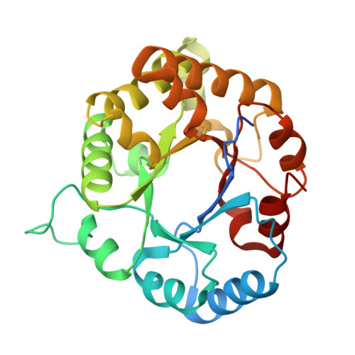
Stable monomers in the ancestral sequence reconstruction of the last opisthokont common ancestor of dimeric triosephosphate isomerase.

Perez-Nino, J.A.Guerra, Y.Diaz-Salazar, A.J.Costas, M.Rodriguez-Romero, A.Fernandez-Velasco, D.A.

(2024) Protein Sci 33: e5134-e5134

  • DOI: https://doi.org/10.1002/pro.5134
  • Primary Citation of Related Structures:  
    8V09, 8V0A, 8V2W, 8V2X, 8W05, 8W06, 8W08

  • PubMed Abstract: 

    Function and structure are strongly coupled in obligated oligomers such as Triosephosphate isomerase (TIM). In animals and fungi, TIM monomers are inactive and unstable. Previously, we used ancestral sequence reconstruction to study TIM evolution and found that before these lineages diverged, the last opisthokonta common ancestor of TIM (LOCATIM) was an obligated oligomer that resembles those of extant TIMs. Notably, calorimetric evidence indicated that ancestral TIM monomers are more structured than extant ones. To further increase confidence about the function, structure, and stability of the LOCATIM, in this work, we applied two different inference methodologies and the worst plausible case scenario for both of them, to infer four sequences of this ancestor and test the robustness of their physicochemical properties. The extensive biophysical characterization of the four reconstructed sequences of LOCATIM showed very similar hydrodynamic and spectroscopic properties, as well as ligand-binding energetics and catalytic parameters. Their 3D structures were also conserved. Although differences were observed in melting temperature, all LOCATIMs showed reversible urea-induced unfolding transitions, and for those that reached equilibrium, high conformational stability was estimated (ΔG Tot  = 40.6-46.2 kcal/mol). The stability of the inactive monomeric intermediates was also high (ΔG unf  = 12.6-18.4 kcal/mol), resembling some protozoan TIMs rather than the unstable monomer observed in extant opisthokonts. A comparative analysis of the 3D structure of ancestral and extant TIMs shows a correlation between the higher stability of the ancestral monomers with the presence of several hydrogen bonds located in the "bottom" part of the barrel.


  • Organizational Affiliation

    Laboratorio de Fisicoquímica e Ingeniería de Proteínas, Departamento de Bioquímica, Facultad de Medicina, Universidad Nacional Autónoma de México, Ciudad de México, Mexico.


Macromolecules
Find similar proteins by:  (by identity cutoff)  |  3D Structure
Entity ID: 1
MoleculeChains Sequence LengthOrganismDetailsImage
Triosephosphate isomerase
A, B, C, D, E
A, B, C, D, E, F, G, H
270synthetic constructMutation(s): 0 
EC: 5.3.1.1
Entity Groups  
Sequence Clusters30% Identity50% Identity70% Identity90% Identity95% Identity100% Identity
Sequence Annotations
Expand
  • Reference Sequence
Small Molecules
Ligands 8 Unique
IDChains Name / Formula / InChI Key2D Diagram3D Interactions
PGH (Subject of Investigation/LOI)
Query on PGH

Download Ideal Coordinates CCD File 
I [auth A]
J [auth B]
L [auth C]
P [auth D]
R [auth E]
I [auth A],
J [auth B],
L [auth C],
P [auth D],
R [auth E],
U [auth F],
V [auth G],
X [auth H]
PHOSPHOGLYCOLOHYDROXAMIC ACID
C2 H6 N O6 P
BAXHHWZKQZIJID-UHFFFAOYSA-N
PGE
Query on PGE

Download Ideal Coordinates CCD File 
M [auth C]TRIETHYLENE GLYCOL
C6 H14 O4
ZIBGPFATKBEMQZ-UHFFFAOYSA-N
PEG
Query on PEG

Download Ideal Coordinates CCD File 
S [auth E],
W [auth G]
DI(HYDROXYETHYL)ETHER
C4 H10 O3
MTHSVFCYNBDYFN-UHFFFAOYSA-N
EDO
Query on EDO

Download Ideal Coordinates CCD File 
Q [auth D]1,2-ETHANEDIOL
C2 H6 O2
LYCAIKOWRPUZTN-UHFFFAOYSA-N
ACY
Query on ACY

Download Ideal Coordinates CCD File 
K [auth B]ACETIC ACID
C2 H4 O2
QTBSBXVTEAMEQO-UHFFFAOYSA-N
ACT
Query on ACT

Download Ideal Coordinates CCD File 
N [auth C]ACETATE ION
C2 H3 O2
QTBSBXVTEAMEQO-UHFFFAOYSA-M
CL
Query on CL

Download Ideal Coordinates CCD File 
T [auth F]CHLORIDE ION
Cl
VEXZGXHMUGYJMC-UHFFFAOYSA-M
NA
Query on NA

Download Ideal Coordinates CCD File 
O [auth D]SODIUM ION
Na
FKNQFGJONOIPTF-UHFFFAOYSA-N
Experimental Data & Validation

Experimental Data

  • Method: X-RAY DIFFRACTION
  • Resolution: 2.06 Å
  • R-Value Free: 0.235 
  • R-Value Work: 0.195 
  • R-Value Observed: 0.197 
  • Space Group: P 1 21 1
Unit Cell:
Length ( Å )Angle ( ˚ )
a = 111.642α = 90
b = 83.222β = 103.49
c = 127.865γ = 90
Software Package:
Software NamePurpose
PHENIXrefinement
HKL-3000data reduction
HKL-3000data scaling
PHASERphasing

Structure Validation

View Full Validation Report


Ligand Structure Quality Assessment 


Entry History & Funding Information

Deposition Data


Funding OrganizationLocationGrant Number
Other governmentMexicoIV200322
Other governmentMexicoIN210519

Revision History  (Full details and data files)

  • Version 1.0: 2024-09-04
    Type: Initial release