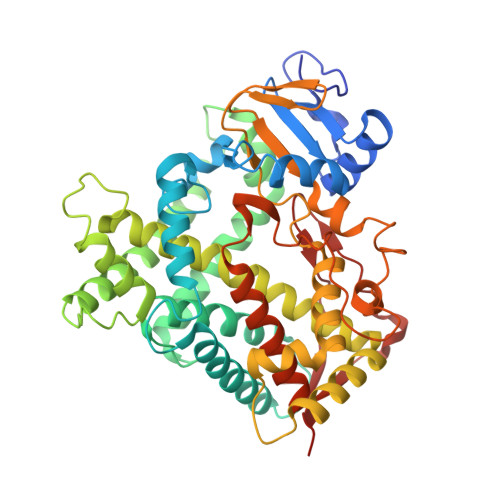
Structural and biophysical analysis of cytochrome P450 2C9*14 and *27 variants in complex with losartan.
Parikh, S.J., Edara, S., Deodhar, S., Singh, A.K., Maekawa, K., Zhang, Q., Glass, K.C., Shah, M.B.(2024) J Inorg Biochem 258: 112622-112622
- PubMed: 38852293
- DOI: https://doi.org/10.1016/j.jinorgbio.2024.112622
- Primary Citation of Related Structures:
8VX0, 8VZ7 - PubMed Abstract:
The human cytochrome P450 (CYP) 1, 2 and 3 families of enzymes are responsible for the biotransformation of a majority of the currently available pharmaceutical drugs. The highly polymorphic CYP2C9 predominantly metabolizes many drugs including anticoagulant S-warfarin, anti-hypertensive losartan, anti-diabetic tolbutamide, analgesic ibuprofen, etc. There are >80 single nucleotide changes identified in CYP2C9, many of which significantly alter the clearance of important drugs. Here we report the structural and biophysical analysis of two polymorphic variants, CYP2C9*14 (Arg125His) and CYP2C9*27 (Arg150Leu) complexed with losartan. The X-ray crystal structures of the CYP2C9*14 and *27 illustrate the binding of two losartan molecules, one in the active site near heme and another on the periphery. Both losartan molecules are bound in an identical conformation to that observed in the previously solved CYP2C9 wild-type complex, however, the number of losartan differs from the wild-type structure, which showed binding of three molecules. Additionally, isothermal titration calorimetry experiments reveal a lower binding affinity of losartan with *14 and *27 variants when compared to the wild-type. Overall, the results provide new insights into the effects of these genetic polymorphisms and suggests a possible mechanism contributing to reduced metabolic activity in patients carrying these alleles.
- Department of Pharmaceutical Sciences, Albany College of Pharmacy and Health Sciences, 106 New Scotland Avenue, Albany, NY 12208, USA.
Organizational Affiliation:


