8VTZ | pdb_00008vtz

Crystal structure of Aquifex aeolicus Trbp111


Experimental Data Snapshot

  • Method: X-RAY DIFFRACTION
  • Resolution: 2.30 Å
  • R-Value Free: 
    0.264 (Depositor), 0.264 (DCC) 
  • R-Value Work: 
    0.214 (Depositor), 0.214 (DCC) 
  • R-Value Observed: 
    0.216 (Depositor) 

Starting Model: experimental
View more details

wwPDB Validation 3D Report Full Report

Validation slider image for 8VTZ

This is version 1.0 of the entry. See complete history

Literature

Structural basis of tRNA recognition by the widespread OB fold.

Umuhire Juru, A.Ghirlando, R.Zhang, J.

(2024) Nat Commun 15: 6385-6385

  • DOI: https://doi.org/10.1038/s41467-024-50730-1
  • Primary Citation Related Structures: 
    8VTZ, 8VU0

  • PubMed Abstract: 

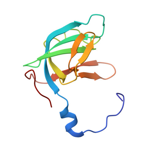
    The widespread oligonucleotide/oligosaccharide-binding (OB)-fold recognizes diverse substrates from sugars to nucleic acids and proteins, and plays key roles in genome maintenance, transcription, translation, and tRNA metabolism. OB-containing bacterial Trbp and yeast Arc1p proteins are thought to recognize the tRNA elbow or anticodon regions. Here we report a 2.6 Å co-crystal structure of Aquifex aeolicus Trbp111 bound to tRNA Ile , which reveals that Trbp recognizes tRNAs solely by capturing their 3' ends. Structural, mutational, and biophysical analyses show that the Trbp/EMAPII-like OB fold precisely recognizes the single-stranded structure, 3' terminal location, and specific sequence of the 3' CA dinucleotide - a universal feature of mature tRNAs. Arc1p supplements its OB - tRNA 3' end interaction with additional contacts that involve an adjacent basic region and the tRNA body. This study uncovers a previously unrecognized mode of tRNA recognition by an ancient protein fold, and provides insights into protein-mediated tRNA aminoacylation, folding, localization, trafficking, and piracy.


  • Organizational Affiliation
    • Laboratory of Molecular Biology, National Institute of Diabetes and Digestive and Kidney Diseases, Bethesda, MD, USA.

Macromolecule Content 

  • Total Structure Weight: 24.63 kDa 
  • Atom Count: 1,779 
  • Modeled Residue Count: 226 
  • Deposited Residue Count: 226 
  • Unique protein chains: 1

Macromolecules

Find similar proteins by:|  3D Structure
Entity ID: 1
MoleculeChains  Sequence LengthOrganismDetailsImage
Methionyl-tRNA synthetase beta subunit
A, B
113Aquifex aeolicusMutation(s): 0 
Gene Names: metG'aq_422
EC: 6.1.1.10
UniProt
Find proteins for O66738 (Aquifex aeolicus (strain VF5))
Explore O66738 
Go to UniProtKB:  O66738
Entity Groups
Sequence Clusters30% Identity50% Identity70% Identity90% Identity95% Identity100% Identity
UniProt GroupO66738
Sequence Annotations
Expand
Reference Sequence

Small Molecules

Ligands 1 Unique
IDChains Name / Formula / InChI Key2D Diagram3D Interactions
SO4

Query on SO4


Download:Ideal Coordinates CCD File
C [auth B]SULFATE ION
O4 S
QAOWNCQODCNURD-UHFFFAOYSA-L

Experimental Data & Validation

Experimental Data

  • Method: X-RAY DIFFRACTION
  • Resolution: 2.30 Å
  • R-Value Free:  0.264 (Depositor), 0.264 (DCC) 
  • R-Value Work:  0.214 (Depositor), 0.214 (DCC) 
  • R-Value Observed: 0.216 (Depositor) 
Space Group: C 2 2 21
Unit Cell:
Length ( Å )Angle ( ˚ )
a = 58.002α = 90
b = 82.221β = 90
c = 88.882γ = 90
Software Package:
Software NamePurpose
PHENIXrefinement
DIALSdata reduction
DIALSdata scaling
PHASERphasing

Structure Validation

View Full Validation Report


Entry History 

& Funding Information

Deposition Data


Funding OrganizationLocationGrant Number
National Institutes of Health/National Institute of Diabetes and Digestive and Kidney Disease (NIH/NIDDK)United StatesZIADK075136

Revision History  (Full details and data files)

  • Version 1.0: 2024-08-07
    Type: Initial release