8VRB

Structure of a synthetic antibody in complex with a class I MHC presenting a hapten-peptide conjugate


Experimental Data Snapshot

  • Method: ELECTRON MICROSCOPY
  • Resolution: 3.25 Å
  • Aggregation State: PARTICLE 
  • Reconstruction Method: SINGLE PARTICLE 

wwPDB Validation   3D Report Full Report


This is version 1.0 of the entry. See complete history


Literature

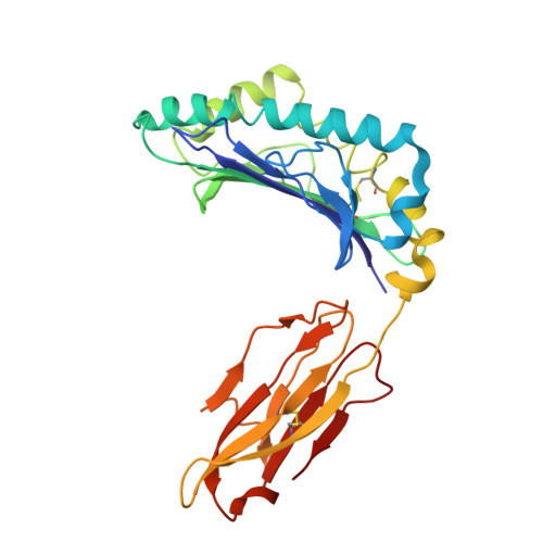
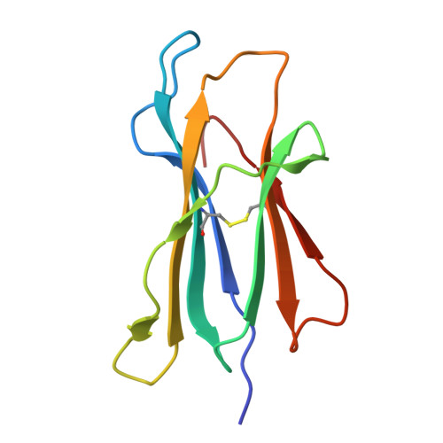
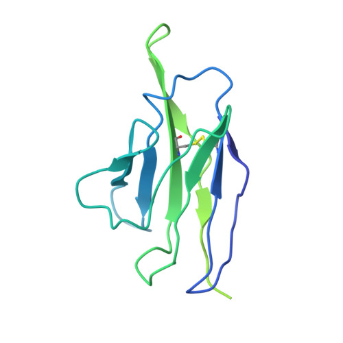
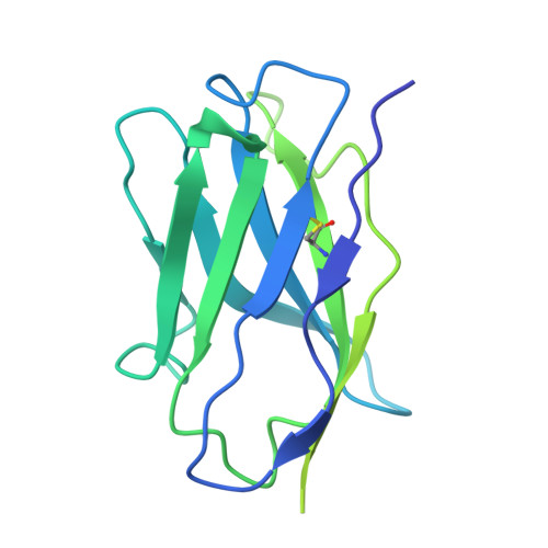
Molecular basis for antibody recognition of multiple drug-peptide/MHC complexes.

Maso, L.Rajak, E.Bang, I.Koide, A.Hattori, T.Neel, B.G.Koide, S.

(2024) Proc Natl Acad Sci U S A 121: e2319029121-e2319029121

  • DOI: https://doi.org/10.1073/pnas.2319029121
  • Primary Citation of Related Structures:  
    8VR9, 8VRA, 8VRB

  • PubMed Abstract: 

    The HapImmune TM platform exploits covalent inhibitors as haptens for creating major histocompatibility complex (MHC)-presented tumor-specific neoantigens by design, combining targeted therapies with immunotherapy for the treatment of drug-resistant cancers. A HapImmune antibody, R023, recognizes multiple sotorasib-conjugated KRAS(G12C) peptides presented by different human leukocyte antigens (HLAs). This high specificity to sotorasib, coupled with broad HLA-binding capability, enables such antibodies, when reformatted as T cell engagers, to potently and selectively kill sotorasib-resistant KRAS(G12C) cancer cells expressing different HLAs upon sotorasib treatment. The loosening of HLA restriction could increase the patient population that can benefit from this therapeutic approach. To understand the molecular basis for its unconventional binding capability, we used single-particle cryogenic electron microscopy to determine the structures of R023 bound to multiple sotorasib-peptide conjugates presented by different HLAs. R023 forms a pocket for sotorasib between the V H and V L domains, binds HLAs in an unconventional, angled way, with V L making most contacts with them, and makes few contacts with the peptide moieties. This binding mode enables the antibody to accommodate different hapten-peptide conjugates and to adjust its conformation to different HLAs presenting hapten-peptides. Deep mutational scanning validated the structures and revealed distinct levels of mutation tolerance by sotorasib- and HLA-binding residues. Together, our structural information and sequence landscape analysis reveal key features for achieving MHC-restricted recognition of multiple hapten-peptide antigens, which will inform the development of next-generation therapeutic antibodies.


  • Organizational Affiliation

    Laura and Isaac Perlmutter Cancer Center, New York University Langone Health, New York, NY 10016.


Macromolecules
Find similar proteins by:  (by identity cutoff)  |  3D Structure
Entity ID: 1
MoleculeChains Sequence LengthOrganismDetailsImage
HLA-A antigen274Homo sapiensMutation(s): 0 
UniProt
Find proteins for A0A3G8GE10 (Homo sapiens)
Explore A0A3G8GE10 
Go to UniProtKB:  A0A3G8GE10
Entity Groups  
Sequence Clusters30% Identity50% Identity70% Identity90% Identity95% Identity100% Identity
UniProt GroupA0A3G8GE10
Sequence Annotations
Expand
  • Reference Sequence
Find similar proteins by:  (by identity cutoff)  |  3D Structure
Entity ID: 2
MoleculeChains Sequence LengthOrganismDetailsImage
Beta-2-microglobulin100Homo sapiensMutation(s): 0 
Gene Names: B2MCDABP0092HDCMA22P
UniProt & NIH Common Fund Data Resources
Find proteins for P61769 (Homo sapiens)
Explore P61769 
Go to UniProtKB:  P61769
PHAROS:  P61769
GTEx:  ENSG00000166710 
Entity Groups  
Sequence Clusters30% Identity50% Identity70% Identity90% Identity95% Identity100% Identity
UniProt GroupP61769
Sequence Annotations
Expand
  • Reference Sequence

Find similar proteins by:  Sequence   |   3D Structure  

Entity ID: 3
MoleculeChains Sequence LengthOrganismDetailsImage
GTPase KRas, N-terminally processed10Homo sapiensMutation(s): 1 
UniProt & NIH Common Fund Data Resources
Find proteins for P01116 (Homo sapiens)
Explore P01116 
Go to UniProtKB:  P01116
PHAROS:  P01116
GTEx:  ENSG00000133703 
Entity Groups  
Sequence Clusters30% Identity50% Identity70% Identity90% Identity95% Identity100% Identity
UniProt GroupP01116
Sequence Annotations
Expand
  • Reference Sequence
Find similar proteins by:  (by identity cutoff)  |  3D Structure
Entity ID: 4
MoleculeChains Sequence LengthOrganismDetailsImage
R023 Fab light chain216Homo sapiensMutation(s): 0 
Entity Groups  
Sequence Clusters30% Identity50% Identity70% Identity90% Identity95% Identity100% Identity
Sequence Annotations
Expand
  • Reference Sequence
Find similar proteins by:  (by identity cutoff)  |  3D Structure
Entity ID: 5
MoleculeChains Sequence LengthOrganismDetailsImage
R023 Fab heavy chain228Homo sapiensMutation(s): 0 
Entity Groups  
Sequence Clusters30% Identity50% Identity70% Identity90% Identity95% Identity100% Identity
Sequence Annotations
Expand
  • Reference Sequence
Small Molecules
Ligands 1 Unique
IDChains Name / Formula / InChI Key2D Diagram3D Interactions
MOV (Subject of Investigation/LOI)
Query on MOV

Download Ideal Coordinates CCD File 
F [auth E]AMG 510 (bound form)
C30 H32 F2 N6 O3
ZIXPBHVQXGLHAG-SFHVURJKSA-N
Experimental Data & Validation

Experimental Data

  • Method: ELECTRON MICROSCOPY
  • Resolution: 3.25 Å
  • Aggregation State: PARTICLE 
  • Reconstruction Method: SINGLE PARTICLE 

Structure Validation

View Full Validation Report


Entry History & Funding Information

Deposition Data


Funding OrganizationLocationGrant Number
National Institutes of Health/National Cancer Institute (NIH/NCI)United States--

Revision History  (Full details and data files)

  • Version 1.0: 2024-09-04
    Type: Initial release