


Crystal Structure of Betaine aldehyde dehydrogenase (BetB) from Klebsiella aerogenes (CoA bound)
Liu, L., Lovell, S., Battaile, K.P., Cooper, A.To be published.
Experimental Data Snapshot
Starting Model: experimental
View more details
Entity ID: 1 | |||||
|---|---|---|---|---|---|
| Molecule | Chains | Sequence Length | Organism | Details | Image |
| Betaine aldehyde dehydrogenase | 499 | Klebsiella aerogenes KCTC 2190 | Mutation(s): 2 Gene Names: betB_3 EC: 1.2.1.8 | 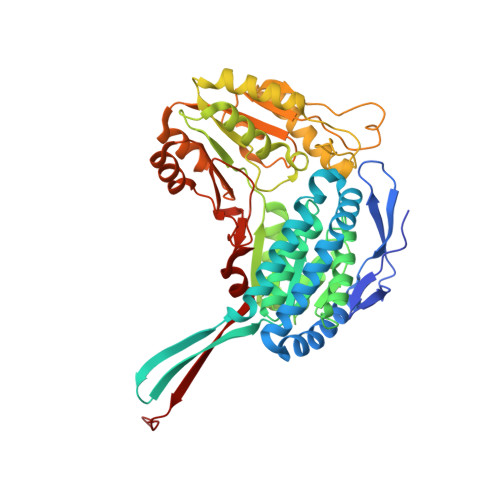 | |
UniProt | |||||
Find proteins for A0A0H3FPU4 (Klebsiella aerogenes (strain ATCC 13048 / DSM 30053 / CCUG 1429 / JCM 1235 / KCTC 2190 / NBRC 13534 / NCIMB 10102 / NCTC 10006 / CDC 819-56)) Explore A0A0H3FPU4 Go to UniProtKB: A0A0H3FPU4 | |||||
Entity Groups | |||||
| Sequence Clusters | 30% Identity50% Identity70% Identity90% Identity95% Identity100% Identity | ||||
| UniProt Group | A0A0H3FPU4 | ||||
Sequence AnnotationsExpand | |||||
Reference Sequence | |||||
| Ligands 5 Unique | |||||
|---|---|---|---|---|---|
| ID | Chains | Name / Formula / InChI Key | 2D Diagram | 3D Interactions | |
| COA (Subject of Investigation/LOI) Download:Ideal Coordinates CCD File | I [auth C], J [auth D] | COENZYME A C21 H36 N7 O16 P3 S RGJOEKWQDUBAIZ-IBOSZNHHSA-N |  | ||
| PGE Download:Ideal Coordinates CCD File | E [auth A] | TRIETHYLENE GLYCOL C6 H14 O4 ZIBGPFATKBEMQZ-UHFFFAOYSA-N |  | ||
| PEG Download:Ideal Coordinates CCD File | G [auth B] | DI(HYDROXYETHYL)ETHER C4 H10 O3 MTHSVFCYNBDYFN-UHFFFAOYSA-N |  | ||
| GOL Download:Ideal Coordinates CCD File | F [auth B] | GLYCEROL C3 H8 O3 PEDCQBHIVMGVHV-UHFFFAOYSA-N |  | ||
| CL Download:Ideal Coordinates CCD File | H [auth B] | CHLORIDE ION Cl VEXZGXHMUGYJMC-UHFFFAOYSA-M |  | ||
| Length ( Å ) | Angle ( ˚ ) |
|---|---|
| a = 83.818 | α = 90 |
| b = 101.008 | β = 95.94 |
| c = 123.928 | γ = 90 |
| Software Name | Purpose |
|---|---|
| PHENIX | refinement |
| Aimless | data scaling |
| XDS | data reduction |
| PHASER | phasing |
| Funding Organization | Location | Grant Number |
|---|---|---|
| National Institutes of Health/National Institute Of Allergy and Infectious Diseases (NIH/NIAID) | United States | 75N93022C00036 |
| National Institutes of Health/Office of the Director | United States | S10OD030394 |


