Allosteric activation of VCP, an AAA unfoldase, by small molecule mimicry.
Jones, N.H., Liu, Q., Urnavicius, L., Dahan, N.E., Vostal, L.E., Kapoor, T.M.(2024) Proc Natl Acad Sci U S A 121: e2316892121-e2316892121
- PubMed: 38833472 Search on PubMedSearch on PubMed Central
- DOI: https://doi.org/10.1073/pnas.2316892121
- Primary Citation Related Structures:
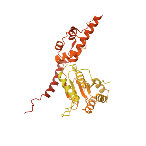
8VKU, 8VLS, 8VOV - PubMed Abstract:
The loss of function of AAA (ATPases associated with diverse cellular activities) mechanoenzymes has been linked to diseases, and small molecules that activate these proteins can be powerful tools to probe mechanisms and test therapeutic hypotheses. Unlike chemical inhibitors that can bind a single conformational state to block enzyme function, activator binding must be permissive to different conformational states needed for mechanochemistry. However, we do not know how AAA proteins can be activated by small molecules. Here, we focus on valosin-containing protein (VCP)/p97, an AAA unfoldase whose loss of function has been linked to protein aggregation-based disorders, to identify druggable sites for chemical activators. We identified VCP ATPase Activator 1 (VAA1), a compound that dose-dependently stimulates VCP ATPase activity up to ~threefold. Our cryo-EM studies resulted in structures (ranging from ~2.9 to 3.7 Å-resolution) of VCP in apo and ADP-bound states and revealed that VAA1 binds an allosteric pocket near the C-terminus in both states. Engineered mutations in the VAA1-binding site confer resistance to VAA1, and furthermore, modulate VCP activity. Mutation of a phenylalanine residue in the VCP C-terminal tail that can occupy the VAA1 binding site also stimulates ATPase activity, suggesting that VAA1 acts by mimicking this interaction. Together, our findings uncover a druggable allosteric site and a mechanism of enzyme regulation that can be tuned through small molecule mimicry.
- Laboratory of Chemistry and Cell Biology, The Rockefeller University, New York, NY 10065.
Organizational Affiliation: