8VKB | pdb_00008vkb

Crystal structure of Plasmodium vivax glycylpeptide N-tetradecanoyltransferase (N-myristoyltransferase, NMT) bound to myristoyl-CoA and inhibitor 10b


Experimental Data Snapshot

  • Method: X-RAY DIFFRACTION
  • Resolution: 2.43 Å
  • R-Value Free: 
    0.233 (Depositor), 0.233 (DCC) 
  • R-Value Work: 
    0.185 (Depositor), 0.185 (DCC) 
  • R-Value Observed: 
    0.187 (Depositor) 

Starting Model: experimental
View more details

wwPDB Validation 3D Report Full Report

Validation slider image for 8VKB

Ligand Structure Quality Assessment 


This is version 1.0 of the entry. See complete history

Literature

Exploring Subsite Selectivity within Plasmodium vivax N -Myristoyltransferase Using Pyrazole-Derived Inhibitors.

Rodriguez-Hernandez, D.Fenwick, M.K.Zigweid, R.Sankaran, B.Myler, P.J.Sunnerhagen, P.Kaushansky, A.Staker, B.L.Grotli, M.

(2024) J Med Chem 67: 7312-7329

  • DOI: https://doi.org/10.1021/acs.jmedchem.4c00168
  • Primary Citation Related Structures: 
    8VKA, 8VKB

  • PubMed Abstract: 

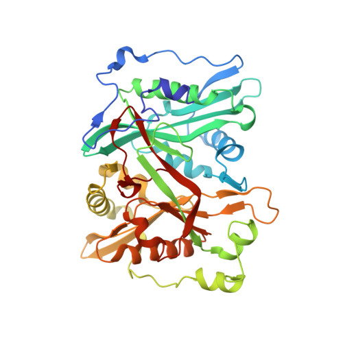
    N -myristoyltransferase (NMT) is a promising antimalarial drug target. Despite biochemical similarities between Plasmodium vivax and human NMTs, our recent research demonstrated that high selectivity is achievable. Herein, we report Pv NMT-inhibiting compounds aimed at identifying novel mechanisms of selectivity. Various functional groups are appended to a pyrazole moiety in the inhibitor to target a pocket formed beneath the peptide binding cleft. The inhibitor core group polarity, lipophilicity, and size are also varied to probe the water structure near a channel. Selectivity index values range from 0.8 to 125.3. Cocrystal structures of two selective compounds, determined at 1.97 and 2.43 Å, show that extensions bind the targeted pocket but with different stabilities. A bulky naphthalene moiety introduced into the core binds next to instead of displacing protein-bound waters, causing a shift in the inhibitor position and expanding the binding site. Our structure-activity data provide a conceptual foundation for guiding future inhibitor optimizations.


  • Organizational Affiliation
    • Department of Chemistry and Molecular Biology, University of Gothenburg, S-405 30 Gothenburg, Sweden.

Macromolecule Content 

  • Total Structure Weight: 141.29 kDa 
  • Atom Count: 10,707 
  • Modeled Residue Count: 1,153 
  • Deposited Residue Count: 1,158 
  • Unique protein chains: 1

Macromolecules

Find similar proteins by:|  3D Structure
Entity ID: 1
MoleculeChains  Sequence LengthOrganismDetailsImage
Glycylpeptide N-tetradecanoyltransferase
A, B, C
386Plasmodium vivax Sal-1Mutation(s): 0 
Gene Names: PVX_085815
EC: 2.3.1.97
UniProt
Find proteins for A5K1A2 (Plasmodium vivax (strain Salvador I))
Explore A5K1A2 
Go to UniProtKB:  A5K1A2
Entity Groups
Sequence Clusters30% Identity50% Identity70% Identity90% Identity95% Identity100% Identity
UniProt GroupA5K1A2
Sequence Annotations
Expand
Reference Sequence

Small Molecules

Ligands 6 Unique
IDChains Name / Formula / InChI Key2D Diagram3D Interactions
MYA
(Subject of Investigation/LOI)

Query on MYA


Download:Ideal Coordinates CCD File
D [auth A],
M [auth B],
V [auth C]
TETRADECANOYL-COA
C35 H62 N7 O17 P3 S
DUAFKXOFBZQTQE-QSGBVPJFSA-N
A1AB8
(Subject of Investigation/LOI)

Query on A1AB8


Download:Ideal Coordinates CCD File
E [auth A],
N [auth B],
W [auth C]
{1,5-dimethyl-4-[2-({(1P)-1-[2-(piperazin-1-yl)pyridin-4-yl]naphthalen-2-yl}oxy)ethyl]-1H-pyrazol-3-yl}(morpholin-4-yl)methanone
C31 H36 N6 O3
DUUNCVRAOLWCAR-UHFFFAOYSA-N
P6G

Query on P6G


Download:Ideal Coordinates CCD File
T [auth B]HEXAETHYLENE GLYCOL
C12 H26 O7
IIRDTKBZINWQAW-UHFFFAOYSA-N
1PE

Query on 1PE


Download:Ideal Coordinates CCD File
J [auth A]PENTAETHYLENE GLYCOL
C10 H22 O6
JLFNLZLINWHATN-UHFFFAOYSA-N
GOL

Query on GOL


Download:Ideal Coordinates CCD File
FA [auth C],
I [auth A],
U [auth B]
GLYCEROL
C3 H8 O3
PEDCQBHIVMGVHV-UHFFFAOYSA-N
CL

Query on CL


Download:Ideal Coordinates CCD File
AA [auth C]
BA [auth C]
CA [auth C]
DA [auth C]
EA [auth C]
AA [auth C],
BA [auth C],
CA [auth C],
DA [auth C],
EA [auth C],
F [auth A],
G [auth A],
H [auth A],
K [auth A],
L [auth A],
O [auth B],
P [auth B],
Q [auth B],
R [auth B],
S [auth B],
X [auth C],
Y [auth C],
Z [auth C]
CHLORIDE ION
Cl
VEXZGXHMUGYJMC-UHFFFAOYSA-M

Experimental Data & Validation

Experimental Data

  • Method: X-RAY DIFFRACTION
  • Resolution: 2.43 Å
  • R-Value Free:  0.233 (Depositor), 0.233 (DCC) 
  • R-Value Work:  0.185 (Depositor), 0.185 (DCC) 
  • R-Value Observed: 0.187 (Depositor) 
Space Group: P 21 21 21
Unit Cell:
Length ( Å )Angle ( ˚ )
a = 57.366α = 90
b = 119.781β = 90
c = 176.883γ = 90
Software Package:
Software NamePurpose
XDSdata reduction
PHENIXrefinement
Aimlessdata scaling
PHASERphasing
Cootmodel building
PDB_EXTRACTdata extraction

Structure Validation

View Full Validation Report


Ligand Structure Quality Assessment 


Entry History 

& Funding Information

Deposition Data


Funding OrganizationLocationGrant Number
National Institutes of Health/National Institute Of Allergy and Infectious Diseases (NIH/NIAID)United StatesR01AI155536
National Institutes of Health/National Institute Of Allergy and Infectious Diseases (NIH/NIAID)United States75N93022C00036

Revision History  (Full details and data files)

  • Version 1.0: 2024-07-17
    Type: Initial release