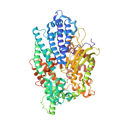
Structural basis of divergent substrate recognition and inhibition of human neurolysin.
Shi, K., Bagchi, S., Bickel, J., Esfahani, S.H., Yin, L., Cheng, T., Karamyan, V.T., Aihara, H.(2024) Sci Rep 14: 18420-18420
- PubMed: 39117724 Search on PubMedSearch on PubMed Central
- DOI: https://doi.org/10.1038/s41598-024-67639-w
- Primary Citation Related Structures:
8VJU, 8VJV, 8VJW, 8VJX, 8VJY - PubMed Abstract:
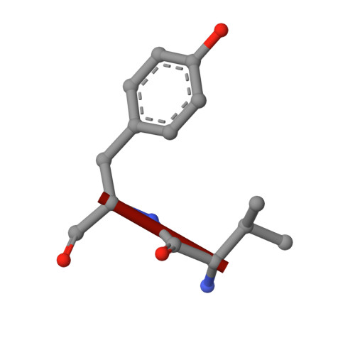
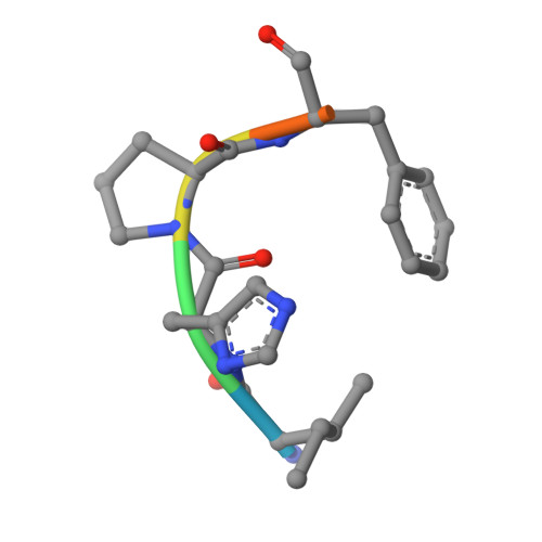
A zinc metallopeptidase neurolysin (Nln) processes diverse bioactive peptides to regulate signaling in the mammalian nervous system. To understand how Nln interacts with various peptides with dissimilar sequences, we determined crystal structures of Nln in complex with diverse peptides including dynorphins, angiotensin, neurotensin, and bradykinin. The structures show that Nln binds these peptides in a large dumbbell-shaped interior cavity constricted at the active site, making minimal structural changes to accommodate different peptide sequences. The structures also show that Nln readily binds similar peptides with distinct registers, which can determine whether the peptide serves as a substrate or a competitive inhibitor. We analyzed the activities and binding of Nln toward various forms of dynorphin A peptides, which highlights the promiscuous nature of peptide binding and shows how dynorphin A (1-13) potently inhibits the Nln activity while dynorphin A (1-8) is efficiently cleaved. Our work provides insights into the broad substrate specificity of Nln and may aid in the future design of small molecule modulators for Nln.
- Department of Biochemistry, Molecular Biology and Biophysics, University of Minnesota, Minneapolis, MN, 55455, USA.
Organizational Affiliation: