8VJC | pdb_00008vjc

Cryo-EM structure of short form insulin receptor (IR-A) with three IGF2 bound, asymmetric conformation.


Experimental Data Snapshot

  • Method: ELECTRON MICROSCOPY
  • Resolution: 3.80 Å
  • Aggregation State: PARTICLE 
  • Reconstruction Method: SINGLE PARTICLE 

wwPDB Validation 3D Report Full Report

Validation slider image for 8VJC

This is version 1.2 of the entry. See complete history

Literature

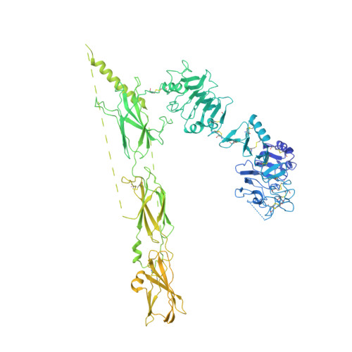
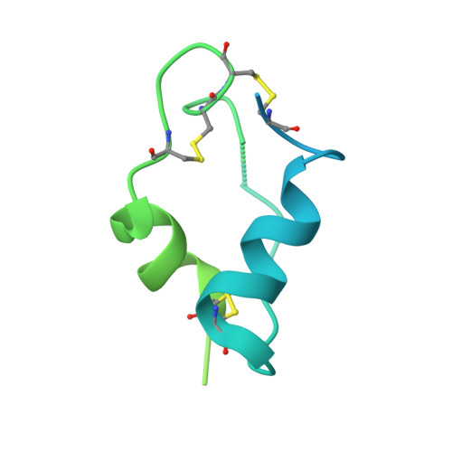
Activation of the insulin receptor by insulin-like growth factor 2.

An, W.Hall, C.Li, J.Hung, A.Wu, J.Park, J.Wang, L.Bai, X.C.Choi, E.

(2024) Nat Commun 15: 2609-2609

  • DOI: https://doi.org/10.1038/s41467-024-46990-6
  • Primary Citation Related Structures: 
    8U4B, 8U4C, 8U4E, 8VJB, 8VJC

  • PubMed Abstract: 

    Insulin receptor (IR) controls growth and metabolism. Insulin-like growth factor 2 (IGF2) has different binding properties on two IR isoforms, mimicking insulin's function. However, the molecular mechanism underlying IGF2-induced IR activation remains unclear. Here, we present cryo-EM structures of full-length human long isoform IR (IR-B) in both the inactive and IGF2-bound active states, and short isoform IR (IR-A) in the IGF2-bound active state. Under saturated IGF2 concentrations, both the IR-A and IR-B adopt predominantly asymmetric conformations with two or three IGF2s bound at site-1 and site-2, which differs from that insulin saturated IR forms an exclusively T-shaped symmetric conformation. IGF2 exhibits a relatively weak binding to IR site-2 compared to insulin, making it less potent in promoting full IR activation. Cell-based experiments validated the functional importance of IGF2 binding to two distinct binding sites in optimal IR signaling and trafficking. In the inactive state, the C-terminus of α-CT of IR-B contacts FnIII-2 domain of the same protomer, hindering its threading into the C-loop of IGF2, thus reducing the association rate of IGF2 with IR-B. Collectively, our studies demonstrate the activation mechanism of IR by IGF2 and reveal the molecular basis underlying the different affinity of IGF2 to IR-A and IR-B.


  • Organizational Affiliation
    • Department of Biophysics, University of Texas Southwestern Medical Center, Dallas, TX, 75390, USA.

Macromolecule Content 

  • Total Structure Weight: 371.17 kDa 
  • Atom Count: 13,927 
  • Modeled Residue Count: 1,726 
  • Deposited Residue Count: 3,280 
  • Unique protein chains: 2

Macromolecules

Find similar proteins by:|  3D Structure
Entity ID: 1
MoleculeChains  Sequence LengthOrganismDetailsImage
Isoform Short of Insulin receptor
A, B
1,370Homo sapiensMutation(s): 0 
Gene Names: INSR
EC: 2.7.10.1
UniProt & NIH Common Fund Data Resources
Find proteins for P06213 (Homo sapiens)
Explore P06213 
Go to UniProtKB:  P06213
PHAROS:  P06213
GTEx:  ENSG00000171105 
Entity Groups
Sequence Clusters30% Identity50% Identity70% Identity90% Identity95% Identity100% Identity
UniProt GroupP06213
Sequence Annotations
Expand
Reference Sequence
Find similar proteins by:|  3D Structure
Entity ID: 2
MoleculeChains  Sequence LengthOrganismDetailsImage
Insulin-like growth factor II
C, D, E
180Homo sapiensMutation(s): 0 
Gene Names: IGF2
UniProt & NIH Common Fund Data Resources
Find proteins for P01344 (Homo sapiens)
Explore P01344 
Go to UniProtKB:  P01344
PHAROS:  P01344
GTEx:  ENSG00000167244 
Entity Groups
Sequence Clusters30% Identity50% Identity70% Identity90% Identity95% Identity100% Identity
UniProt GroupP01344
Sequence Annotations
Expand
Reference Sequence

Experimental Data & Validation

Experimental Data

  • Method: ELECTRON MICROSCOPY
  • Resolution: 3.80 Å
  • Aggregation State: PARTICLE 
  • Reconstruction Method: SINGLE PARTICLE 
EM Software:
TaskSoftware PackageVersion
MODEL REFINEMENTPHENIX
RECONSTRUCTIONRELION

Structure Validation

View Full Validation Report


Entry History 

& Funding Information

Deposition Data


Funding OrganizationLocationGrant Number
National Institutes of Health/National Institute of General Medical Sciences (NIH/NIGMS)United StatesGM136976

Revision History  (Full details and data files)

  • Version 1.0: 2024-03-27
    Type: Initial release
  • Version 1.1: 2024-04-03
    Changes: Database references
  • Version 1.2: 2024-10-23
    Changes: Data collection, Structure summary