Revealing the atomic and electronic mechanism of human manganese superoxide dismutase product inhibition.
Azadmanesh, J., Slobodnik, K., Struble, L.R., Lutz, W.E., Coates, L., Weiss, K.L., Myles, D.A.A., Kroll, T., Borgstahl, G.E.O.(2024) Nat Commun 15: 5973-5973
- PubMed: 39013847 Search on PubMedSearch on PubMed Central
- DOI: https://doi.org/10.1038/s41467-024-50260-w
- Primary Citation Related Structures:
8VHW, 8VHY, 8VJ0, 8VJ4, 8VJ5, 8VJ8 - PubMed Abstract:
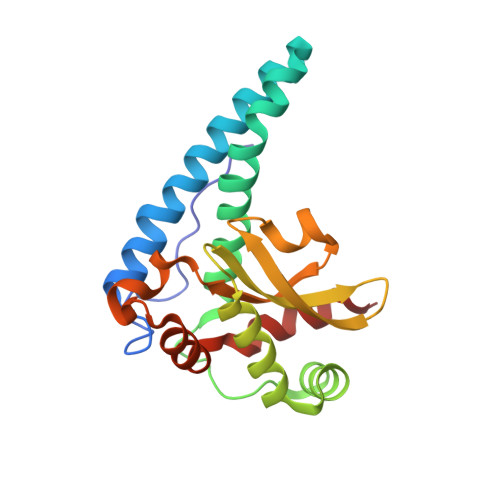
Human manganese superoxide dismutase (MnSOD) is a crucial oxidoreductase that maintains the vitality of mitochondria by converting superoxide (O 2 ●- ) to molecular oxygen (O 2 ) and hydrogen peroxide (H 2 O 2 ) with proton-coupled electron transfers (PCETs). Human MnSOD has evolved to be highly product inhibited to limit the formation of H 2 O 2 , a freely diffusible oxidant and signaling molecule. The product-inhibited complex is thought to be composed of a peroxide (O 2 2- ) or hydroperoxide (HO 2 - ) species bound to Mn ion and formed from an unknown PCET mechanism. PCET mechanisms of proteins are typically not known due to difficulties in detecting the protonation states of specific residues that coincide with the electronic state of the redox center. To shed light on the mechanism, we combine neutron diffraction and X-ray absorption spectroscopy of the product-bound, trivalent, and divalent states of the enzyme to reveal the positions of all the atoms, including hydrogen, and the electronic configuration of the metal ion. The data identifies the product-inhibited complex, and a PCET mechanism of inhibition is constructed.
- Eppley Institute for Research in Cancer and Allied Diseases, 986805 Nebraska Medical Center, Omaha, NE, 68198-6805, USA.
Organizational Affiliation: