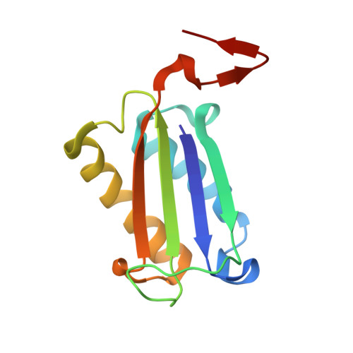
8VFO

Crystal Structure of L117G Variant of D-Dopachrome Tautomerase (D-DT)


Experimental Data Snapshot

  • Method: X-RAY DIFFRACTION
  • Resolution: 1.35 Å
  • R-Value Free: 0.193 
  • R-Value Work: 0.166 
  • R-Value Observed: 0.167 

Starting Model: experimental
View more details

wwPDB Validation   3D Report Full Report


This is version 1.2 of the entry. See complete history


Literature

The C-terminal Region of D-DT Regulates Molecular Recognition for Protein-Ligand Complexes.

Parkins, A.Pilien, A.V.R.Wolff, A.M.Argueta, C.Vargas, J.Sadeghi, S.Franz, A.H.Thompson, M.C.Pantouris, G.

(2024) J Med Chem 67: 7359-7372

  • DOI: https://doi.org/10.1021/acs.jmedchem.4c00177
  • Primary Citation of Related Structures:  
    8VDY, 8VFK, 8VFL, 8VFN, 8VFO, 8VFW, 8VG5, 8VG7, 8VG8

  • PubMed Abstract: 

    Systematic analysis of molecular recognition is critical for understanding the biological function of macromolecules. For the immunomodulatory protein D-dopachrome tautomerase (D-DT), the mechanism of protein-ligand interactions is poorly understood. Here, 17 carefully designed protein variants and wild type (WT) D-DT were interrogated with an array of complementary techniques to elucidate the structural basis of ligand recognition. Utilization of a substrate and two selective inhibitors with distinct binding profiles offered previously unseen mechanistic insights into D-DT-ligand interactions. Our results demonstrate that the C-terminal region serves a key role in molecular recognition via regulation of the active site opening, protein-ligand interactions, and conformational flexibility of the pocket's environment. While our study is the first comprehensive analysis of molecular recognition for D-DT, the findings reported herein promote the understanding of protein functionality and enable the design of new structure-based drug discovery projects.


  • Organizational Affiliation

    Department of Chemistry, University of the Pacific, Stockton, California 95211, United States.


Macromolecules
Find similar proteins by:  (by identity cutoff)  |  3D Structure
Entity ID: 1
MoleculeChains Sequence LengthOrganismDetailsImage
D-dopachrome decarboxylase117Homo sapiensMutation(s): 1 
Gene Names: DDT
EC: 4.1.1.84
UniProt & NIH Common Fund Data Resources
Find proteins for P30046 (Homo sapiens)
Explore P30046 
Go to UniProtKB:  P30046
PHAROS:  P30046
GTEx:  ENSG00000099977 
Entity Groups  
Sequence Clusters30% Identity50% Identity70% Identity90% Identity95% Identity100% Identity
UniProt GroupP30046
Sequence Annotations
Expand
  • Reference Sequence
Small Molecules
Ligands 1 Unique
IDChains Name / Formula / InChI Key2D Diagram3D Interactions
CIT
Query on CIT

Download Ideal Coordinates CCD File 
B [auth A]CITRIC ACID
C6 H8 O7
KRKNYBCHXYNGOX-UHFFFAOYSA-N
Experimental Data & Validation

Experimental Data

  • Method: X-RAY DIFFRACTION
  • Resolution: 1.35 Å
  • R-Value Free: 0.193 
  • R-Value Work: 0.166 
  • R-Value Observed: 0.167 
  • Space Group: H 3
Unit Cell:
Length ( Å )Angle ( ˚ )
a = 80.588α = 90
b = 80.588β = 90
c = 40.123γ = 120
Software Package:
Software NamePurpose
REFMACrefinement
xia2data scaling
MOLREPphasing
XDSdata reduction

Structure Validation

View Full Validation Report


Entry History & Funding Information

Deposition Data


Funding OrganizationLocationGrant Number
Not funded--

Revision History  (Full details and data files)

  • Version 1.0: 2024-05-01
    Type: Initial release
  • Version 1.1: 2024-05-08
    Changes: Database references
  • Version 1.2: 2024-05-22
    Changes: Database references