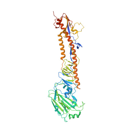
Avian influenza A virus (IAV) surveillance in Northern California, USA, revealed unique IAV hemagglutinin (HA) genome sequences in cloacal swabs from lesser scaups. We found two closely related HA sequences in the same duck species in 2010 and 2013. Phylogenetic analyses suggest that both sequences belong to the recently discovered H19 subtype, which thus far has remained uncharacterized. We demonstrate that H19 does not bind the canonical IAV receptor sialic acid (Sia). Instead, H19 binds to the major histocompatibility complex class II (MHC class II), which facilitates viral entry. Unlike the broad MHC class II specificity of H17 and H18 from bat IAV, H19 exhibits a species-specific MHC class II usage that suggests a limited host range and zoonotic potential. Using cell lines overexpressing MHC class II, we rescued recombinant H19 IAV. We solved the H19 crystal structure and identified residues within the putative Sia receptor binding site (RBS) that impede Sia-dependent entry.
Organizational Affiliation:
Department of Microbiology, Icahn School of Medicine at Mount Sinai, New York, NY 10029, USA; Global Health and Emerging Pathogens Institute, Icahn School of Medicine at Mount Sinai, New York, NY 10029, USA. Electronic address: umut.karakus@mssm.edu.
Department of Microbiology, Icahn School of Medicine at Mount Sinai, New York, NY 10029, USA; Global Health and Emerging Pathogens Institute, Icahn School of Medicine at Mount Sinai, New York, NY 10029, USA; Department of Immunology and Microbiology, The Scripps Research Institute, La Jolla, San Diego, CA 92037, USA.
Departments of Pharmacological Sciences and Oncological Sciences, Tisch Cancer Institute, Icahn School of Medicine at Mount Sinai, New York, NY 10029, USA.
Department of Microbiology, Icahn School of Medicine at Mount Sinai, New York, NY 10029, USA; Global Health and Emerging Pathogens Institute, Icahn School of Medicine at Mount Sinai, New York, NY 10029, USA.
Department of Biology, Barnard College, New York, NY 10027, USA.
Department of Pathology, Microbiology, and Immunology, University of California Davis School of Veterinary Medicine, Davis, CA 95616, USA.
The J. Craig Venter Institute, Rockville, MD 20850, USA.
Department of Chemical Biology & Drug Discovery, Utrecht Institute for Pharmaceutical Sciences, Utrecht University, 3584 CG Utrecht, the Netherlands; Complex Carbohydrate Research Center, University of Georgia, 315 Riverbend Rd, Athens, GA 30602, USA; Bijvoet Center for Biomolecular Research, Utrecht University, 3584 CH Utrecht, the Netherlands; Department of Chemistry, University of Georgia, Athens, GA 30602, USA.
Department of Microbiology, Icahn School of Medicine at Mount Sinai, New York, NY 10029, USA; Center for Vaccine Research and Pandemic Preparedness (C-VaRPP), Icahn School of Medicine at Mount Sinai, New York, NY 10029, USA.
Institute of Medical Virology, University of Zurich, 8057 Zurich, Switzerland.
Department of Chemical Biology & Drug Discovery, Utrecht Institute for Pharmaceutical Sciences, Utrecht University, 3584 CG Utrecht, the Netherlands.
Department of Microbiology, Icahn School of Medicine at Mount Sinai, New York, NY 10029, USA; Global Health and Emerging Pathogens Institute, Icahn School of Medicine at Mount Sinai, New York, NY 10029, USA; Department of Medicine, Division of Infectious Diseases, Icahn School of Medicine at Mount Sinai, New York, NY 10029, USA; The Tisch Cancer Institute, Icahn School of Medicine at Mount Sinai, New York, NY 10029, USA; Department of Pathology, Molecular and Cell-Based Medicine, Icahn School of Medicine at Mount Sinai, New York, NY 10029, USA. Electronic address: adolfo.garcia-sastre@mssm.edu.