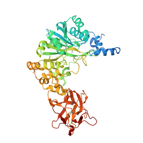
8V9Q

Crystal structure of mGalNAc-T1 in complex with the mucin glycopeptide Muc5AC-13, Mn2+, and UDP.


Experimental Data Snapshot

  • Method: X-RAY DIFFRACTION
  • Resolution: 2.29 Å
  • R-Value Free: 0.278 
  • R-Value Work: 0.248 
  • R-Value Observed: 0.249 

Starting Model: experimental
View more details

wwPDB Validation   3D Report Full Report


Ligand Structure Quality Assessment 


This is version 1.1 of the entry. See complete history


Literature

An unusual dual sugar-binding lectin domain controls the substrate specificity of a mucin-type O-glycosyltransferase.

Collette, A.M.Hassan, S.A.Schmidt, S.I.Lara, A.J.Yang, W.Samara, N.L.

(2024) Sci Adv 10: eadj8829-eadj8829

  • DOI: https://doi.org/10.1126/sciadv.adj8829
  • Primary Citation of Related Structures:  
    8V9Q

  • PubMed Abstract: 

    N-acetylgalactosaminyl-transferases (GalNAc-Ts) initiate mucin-type O-glycosylation, an abundant and complex posttranslational modification that regulates host-microbe interactions, tissue development, and metabolism. GalNAc-Ts contain a lectin domain consisting of three homologous repeats (α, β, and γ), where α and β can potentially interact with O-GalNAc on substrates to enhance activity toward a nearby acceptor Thr/Ser. The ubiquitous isoenzyme GalNAc-T1 modulates heart development, immunity, and SARS-CoV-2 infectivity, but its substrates are largely unknown. Here, we show that both α and β in GalNAc-T1 uniquely orchestrate the O-glycosylation of various glycopeptide substrates. The α repeat directs O-glycosylation to acceptor sites carboxyl-terminal to an existing GalNAc, while the β repeat directs O-glycosylation to amino-terminal sites. In addition, GalNAc-T1 incorporates α and β into various substrate binding modes to cooperatively increase the specificity toward an acceptor site located between two existing O-glycans. Our studies highlight a unique mechanism by which dual lectin repeats expand substrate specificity and provide crucial information for identifying the biological substrates of GalNAc-T1.


  • Organizational Affiliation

    Structural Biochemistry Unit, NIDCR, NIH, Bethesda, MD 20892, USA.


Macromolecules
Find similar proteins by:  (by identity cutoff)  |  3D Structure
Entity ID: 1
MoleculeChains Sequence LengthOrganismDetailsImage
Polypeptide N-acetylgalactosaminyltransferase 1 soluble formA,
C [auth B]
559Mus musculusMutation(s): 0 
Gene Names: Galnt1
EC: 2.4.1.41
UniProt
Find proteins for O08912 (Mus musculus)
Explore O08912 
Go to UniProtKB:  O08912
Entity Groups  
Sequence Clusters30% Identity50% Identity70% Identity90% Identity95% Identity100% Identity
UniProt GroupO08912
Glycosylation
Glycosylation Sites: 2Go to GlyGen: O08912-1
Sequence Annotations
Expand
  • Reference Sequence

Find similar proteins by:  Sequence   |   3D Structure  

Entity ID: 2
MoleculeChains Sequence LengthOrganismDetailsImage
Mucin-5ACB [auth D],
D [auth F],
E [auth H]
16Homo sapiensMutation(s): 0 
UniProt & NIH Common Fund Data Resources
Find proteins for P98088 (Homo sapiens)
Explore P98088 
Go to UniProtKB:  P98088
PHAROS:  P98088
GTEx:  ENSG00000215182 
Entity Groups  
Sequence Clusters30% Identity50% Identity70% Identity90% Identity95% Identity100% Identity
UniProt GroupP98088
Glycosylation
Glycosylation Sites: 1Go to GlyGen: P98088-1
Sequence Annotations
Expand
  • Reference Sequence
Oligosaccharides

Help

Entity ID: 3
MoleculeChains Length2D Diagram Glycosylation3D Interactions
alpha-D-mannopyranose-(1-3)-[alpha-D-mannopyranose-(1-6)]beta-D-mannopyranose-(1-4)-2-acetamido-2-deoxy-beta-D-glucopyranose-(1-4)-2-acetamido-2-deoxy-beta-D-glucopyranoseF [auth X]5N-Glycosylation
Glycosylation Resources
GlyTouCan:  G22768VO
GlyCosmos:  G22768VO
GlyGen:  G22768VO
Entity ID: 4
MoleculeChains Length2D Diagram Glycosylation3D Interactions
2-acetamido-2-deoxy-beta-D-glucopyranose-(1-4)-2-acetamido-2-deoxy-beta-D-glucopyranoseG [auth C]2N-Glycosylation
Glycosylation Resources
GlyTouCan:  G42666HT
GlyCosmos:  G42666HT
GlyGen:  G42666HT
Small Molecules
Ligands 7 Unique
IDChains Name / Formula / InChI Key2D Diagram3D Interactions
UDP (Subject of Investigation/LOI)
Query on UDP

Download Ideal Coordinates CCD File 
H [auth A],
S [auth B]
URIDINE-5'-DIPHOSPHATE
C9 H14 N2 O12 P2
XCCTYIAWTASOJW-XVFCMESISA-N
NAG
Query on NAG

Download Ideal Coordinates CCD File 
T [auth B],
U [auth B]
2-acetamido-2-deoxy-beta-D-glucopyranose
C8 H15 N O6
OVRNDRQMDRJTHS-FMDGEEDCSA-N
A2G (Subject of Investigation/LOI)
Query on A2G

Download Ideal Coordinates CCD File 
P [auth D],
W [auth F],
X [auth H]
2-acetamido-2-deoxy-alpha-D-galactopyranose
C8 H15 N O6
OVRNDRQMDRJTHS-CBQIKETKSA-N
SIN
Query on SIN

Download Ideal Coordinates CCD File 
J [auth A]SUCCINIC ACID
C4 H6 O4
KDYFGRWQOYBRFD-UHFFFAOYSA-N
GOL
Query on GOL

Download Ideal Coordinates CCD File 
I [auth A],
O [auth A]
GLYCEROL
C3 H8 O3
PEDCQBHIVMGVHV-UHFFFAOYSA-N
EDO
Query on EDO

Download Ideal Coordinates CCD File 
K [auth A],
L [auth A],
M [auth A],
Q [auth D],
R [auth D]
1,2-ETHANEDIOL
C2 H6 O2
LYCAIKOWRPUZTN-UHFFFAOYSA-N
MN (Subject of Investigation/LOI)
Query on MN

Download Ideal Coordinates CCD File 
N [auth A],
V [auth B]
MANGANESE (II) ION
Mn
WAEMQWOKJMHJLA-UHFFFAOYSA-N
Experimental Data & Validation

Experimental Data

Unit Cell:
Length ( Å )Angle ( ˚ )
a = 59.635α = 90
b = 72.761β = 95.44
c = 148.498γ = 90
Software Package:
Software NamePurpose
PHENIXrefinement
SERGUIdata collection
HKL-2000data reduction
Aimlessdata scaling
MOLREPphasing

Structure Validation

View Full Validation Report


Ligand Structure Quality Assessment 


Entry History & Funding Information

Deposition Data


Funding OrganizationLocationGrant Number
National Institutes of Health/National Institute of Dental and Craniofacial Research (NIH/NIDCR)United States1-ZIA-DE000754-03

Revision History  (Full details and data files)

  • Version 1.0: 2024-03-13
    Type: Initial release
  • Version 1.1: 2024-10-23
    Changes: Structure summary