Biochemical and structural characterization of a reactive intermediate deaminase A homolog from Streptococcus sanguinis.
Benedict, A.B., Aquino, A.I., Buckner, B.A., Aribam, S.D., Ganguly, C., Thomas, L.M., Bourne, P., Rajan, R., Downs, D.M., Somalinga, V.(2025) Sci Rep 15: 22017-22017
- PubMed: 40596262 Search on PubMedSearch on PubMed Central
- DOI: https://doi.org/10.1038/s41598-025-05264-x
- Primary Citation Related Structures:
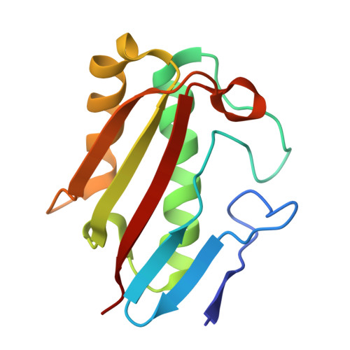
8V8R - PubMed Abstract:
2-Aminoacrylate (2AA) is a short-lived enamine generated as a catalytic intermediate in the dehydration of serine by serine/threonine dehydratase enzymes. 2AA is a metabolic stressor capable of inactivating important pyridoxal phosphate dependent enzymes in a cell. Detoxification of 2AA in a cell is catalyzed by members of the Reactive intermediate deaminase (Rid) family of proteins, which is conserved across all domains of life. We recently identified a RidA homolog, SSA_0809, hereafter SsRidA, in Streptococcus sanguinis with 50% protein sequence identity to a RidA from Salmonella enterica. 2AA deaminase activity assay revealed that SsRidA is capable of enzymatic deamination of 2AA to pyruvate. Furthermore, L-amino acid oxidase assays showed SsRidA has significant activity against several with imino-amino acids similar to the S. enterica RidA. In addition, functional complementation analysis found that SsRidA restored growth of S. enterica ridA mutant in minimal media constituted to increase 2AA stress in the cell. Finally, the crystal structure of SsRidA revealed a homotrimeric protein with active sites at the interface of two interacting monomers. Structure analysis also showed the presence of active site arginine residue along with an active site water molecule implicated in catalysis.
- Department of Biological and Biomedical Sciences, Southwestern Oklahoma State University, Weatherford, OK, USA.
Organizational Affiliation: