Structural Rationalization of IPMK Inhibitor Potency.
Wang, H., Shears, S.B., Blind, R.D.(2025) J Med Chem 68: 24316-24325
- PubMed: 41237254 Search on PubMed
- DOI: https://doi.org/10.1021/acs.jmedchem.5c02314
- Primary Citation Related Structures:
8V6W, 8V6X, 8V6Y, 8V6Z, 8V70, 8V71, 8V72, 8V73, 8V74, 8V75, 8V76, 8V77, 8V78, 8V79 - PubMed Abstract:
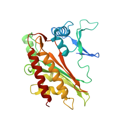
Inositol polyphosphate multikinase (IPMK) is a kinase linked to several cancers; recent development of a large panel of ATP-competitive inhibitors has reinvigorated enthusiasm for targeting IPMK. However, the structural basis for how these inhibitors achieve high potency is unknown. Herein, we report 14 novel cocrystal structures (1.7-2.0 resolution) of human IPMK kinase domain with these inhibitors. We also apply a radiolabeled assay and isothermal titration calorimetry that permit high-confidence IC 50 and K D value determinations. The structures reveal a pocket in the ATP-binding site engaged by the most potent inhibitors. Two ordered waters also participate in hydrogen-bonding networks associated with the most potent inhibitors. In addition to providing the molecular basis for observed increases in potency and selectivity, the data presented here provide a toolbelt of 14 novel inhibitor-bound structures of human IPMK that can serve as a reference for all future IPMK structure-based inhibitor development efforts.
- Molecular and Cellular Biology Laboratory, National Institute of Environmental Health Sciences, NIH, Research Triangle Park, North Carolina 27709, United States.
Organizational Affiliation: