8V5T

Crystal structure of Alzheimers disease phospholipase D3


Experimental Data Snapshot

  • Method: X-RAY DIFFRACTION
  • Resolution: 2.30 Å
  • R-Value Free: 0.209 
  • R-Value Work: 0.185 
  • R-Value Observed: 0.186 

Starting Model: in silico
View more details

wwPDB Validation   3D Report Full Report


This is version 1.0 of the entry. See complete history


Literature

Crystal structure of Alzheimer's disease phospholipase D3 provides a molecular basis for understanding its normal and pathological functions.

Ishii, K.Hermans, S.J.Georgopoulou, M.E.Nero, T.L.Hancock, N.C.Crespi, G.A.N.Gorman, M.A.Gooi, J.H.Parker, M.W.

(2024) FEBS J 

  • DOI: https://doi.org/10.1111/febs.17277
  • Primary Citation of Related Structures:  
    8V5T

  • PubMed Abstract: 

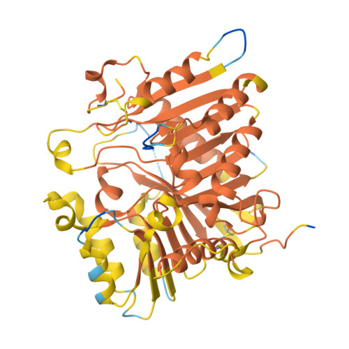
    Human 5'-3' exonuclease PLD3, a member of the phospholipase D family of enzymes, has been validated as a therapeutic target for treating Alzheimer's disease. Here, we have determined the crystal structure of the luminal domain of the enzyme at 2.3 Å resolution, revealing a bilobal structure with a catalytic site located between the lobes. We then compared the structure with published crystal structures of other human PLD family members which revealed that a number of catalytic and lipid recognition residues, previously shown to be key for phospholipase activity, are not conserved or, are absent. This led us to test whether the enzyme is actually a phospholipase. We could not measure any phospholipase activity but the enzyme shows robust nuclease activity. Finally, we have mapped key single nucleotide polymorphisms onto the structure which reveals plausible reasons as to why they have an impact on Alzheimer's disease.


  • Organizational Affiliation

    Structural Biology Laboratory, St Vincent's Institute of Medical Research, Fitzroy, Australia.


Macromolecules
Find similar proteins by:  (by identity cutoff)  |  3D Structure
Entity ID: 1
MoleculeChains Sequence LengthOrganismDetailsImage
5'-3' exonuclease PLD3
A, B
490Homo sapiensMutation(s): 0 
Gene Names: PLD3
UniProt & NIH Common Fund Data Resources
Find proteins for Q8IV08 (Homo sapiens)
Explore Q8IV08 
Go to UniProtKB:  Q8IV08
PHAROS:  Q8IV08
GTEx:  ENSG00000105223 
Entity Groups  
Sequence Clusters30% Identity50% Identity70% Identity90% Identity95% Identity100% Identity
UniProt GroupQ8IV08
Glycosylation
Glycosylation Sites: 2Go to GlyGen: Q8IV08-1
Sequence Annotations
Expand
  • Reference Sequence
Oligosaccharides

Help

Entity ID: 2
MoleculeChains Length2D Diagram Glycosylation3D Interactions
2-acetamido-2-deoxy-beta-D-glucopyranose-(1-4)-2-acetamido-2-deoxy-beta-D-glucopyranose
C, D, E, F
2N-Glycosylation
Small Molecules
Ligands 2 Unique
IDChains Name / Formula / InChI Key2D Diagram3D Interactions
SO4
Query on SO4

Download Ideal Coordinates CCD File 
G [auth A]
H [auth A]
I [auth A]
J [auth A]
K [auth A]
G [auth A],
H [auth A],
I [auth A],
J [auth A],
K [auth A],
L [auth A],
P [auth B],
Q [auth B],
R [auth B],
S [auth B]
SULFATE ION
O4 S
QAOWNCQODCNURD-UHFFFAOYSA-L
GOL
Query on GOL

Download Ideal Coordinates CCD File 
M [auth A],
N [auth A],
O [auth A]
GLYCEROL
C3 H8 O3
PEDCQBHIVMGVHV-UHFFFAOYSA-N
Experimental Data & Validation

Experimental Data

  • Method: X-RAY DIFFRACTION
  • Resolution: 2.30 Å
  • R-Value Free: 0.209 
  • R-Value Work: 0.185 
  • R-Value Observed: 0.186 
  • Space Group: C 1 2 1
Unit Cell:
Length ( Å )Angle ( ˚ )
a = 205.137α = 90
b = 60.566β = 98.72
c = 111.501γ = 90
Software Package:
Software NamePurpose
PHENIXrefinement
XDSdata reduction
Aimlessdata scaling
PHASERphasing

Structure Validation

View Full Validation Report


Entry History & Funding Information

Deposition Data


Funding OrganizationLocationGrant Number
Not funded--

Revision History  (Full details and data files)

  • Version 1.0: 2024-10-09
    Type: Initial release