Structural and functional characterization of the IpaD pi-helix reveals critical roles in DOC interaction, T3SS apparatus maturation, and Shigella virulence.
Barker, S.A., Bernard, A.R., Morales, Y., Johnson, S.J., Dickenson, N.E.(2024) J Biol Chem 300: 107613-107613
- PubMed: 39079629 Search on PubMed
- DOI: https://doi.org/10.1016/j.jbc.2024.107613
- Primary Citation Related Structures:
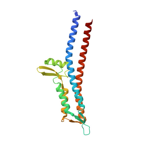
8V5C, 8V5E, 8V7Q, 8V7S - PubMed Abstract:
Shigella spp. are highly pathogenic members of the Enterobacteriaceae family, causing ∼269 million cases of bacillary dysentery and >200,000 deaths each year. Like many Gram-negative pathogens, Shigella rely on their type three secretion system (T3SS) to inject effector proteins into eukaryotic host cells, driving both cellular invasion and evasion of host immune responses. Exposure to the bile salt deoxycholate (DOC) significantly enhances Shigella virulence and is proposed to serve as a critical environmental signal present in the small intestine that prepares Shigella's T3SS for efficient infection of the colonic epithelium. Here, we uncover critical mechanistic details of the Shigella-specific DOC signaling process by describing the role of a π-helix secondary structure element within the T3SS tip protein IpaD. Biophysical characterization and high-resolution structures of IpaD mutants lacking the π-helix show that it is not required for global protein structure, but that it defines the native DOC binding site and prevents off target interactions. Additionally, Shigella strains expressing the π-helix deletion mutants illustrate the pathogenic importance of its role in guiding DOC interaction as flow cytometry and gentamycin protection assays show that the IpaD π-helix is essential for DOC-mediated apparatus maturation and enhanced invasion of eukaryotic cells. Together, these findings add to our understanding of the complex Shigella pathogenesis pathway and its evolution to respond to environmental bile salts by identifying the π-helix in IpaD as a critical structural element required for translating DOC exposure to virulence enhancement.
- From the Department of Chemistry and Biochemistry, Utah State University, Logan, Utah.
Organizational Affiliation: