8UWL

5-HT2AR bound to Lisuride in complex with a mini-Gq protein and an active-state stabilizing single-chain variable fragment (scFv16) obtained by cryo-electron microscopy (cryoEM)


Experimental Data Snapshot

  • Method: ELECTRON MICROSCOPY
  • Resolution: 2.80 Å
  • Aggregation State: PARTICLE 
  • Reconstruction Method: SINGLE PARTICLE 

wwPDB Validation   3D Report Full Report


This is version 1.0 of the entry. See complete history


Literature

AlphaFold2 structures guide prospective ligand discovery.

Lyu, J.Kapolka, N.Gumpper, R.Alon, A.Wang, L.Jain, M.K.Barros-Alvarez, X.Sakamoto, K.Kim, Y.DiBerto, J.Kim, K.Glenn, I.S.Tummino, T.A.Huang, S.Irwin, J.J.Tarkhanova, O.O.Moroz, Y.Skiniotis, G.Kruse, A.C.Shoichet, B.K.Roth, B.L.

(2024) Science : eadn6354-eadn6354

  • DOI: https://doi.org/10.1126/science.adn6354
  • Primary Citation of Related Structures:  
    8UWL, 8V6U

  • PubMed Abstract: 

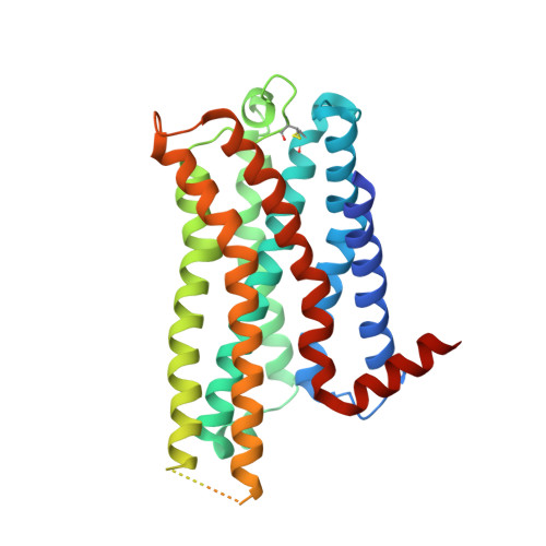
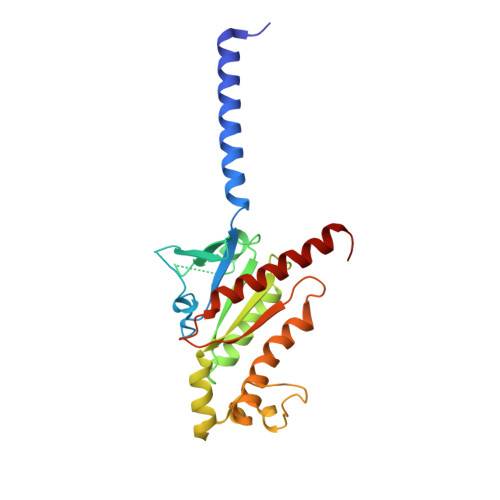
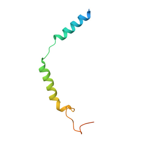
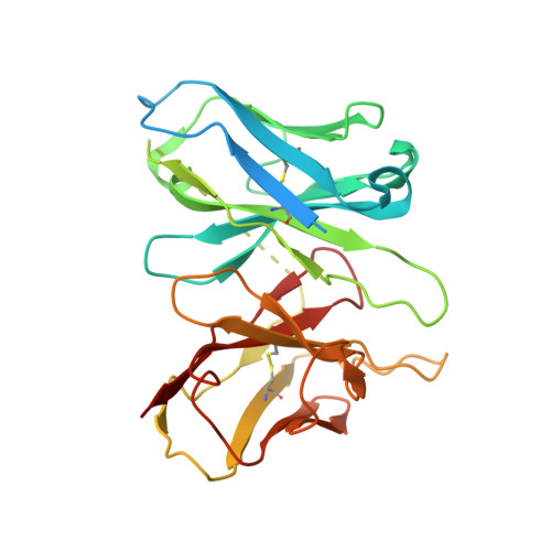
    AlphaFold2 (AF2) models have had wide impact, but they have had mixed success in retrospective ligand recognition. We prospectively docked large libraries against unrefined AF2 models of the σ2 and 5-HT2A receptors, testing hundreds of new molecules and comparing results to docking against the experimental structures. Hit rates were high and similar for the experimental and the AF2 structures, as were affinities. The success of docking against the AF2 models was achieved despite differences in orthosteric residue conformations versus the experimental structures. Determination of the cryo-electron microscopy structure for one of the more potent 5HT2A ligands from the AF2 docking revealed residue accommodations that resembled the AF2 prediction. AF2 models may sample conformations that differ from experimental structures but remain low energy and relevant for ligand discovery, extending the domain of structure-based ligand discovery.


  • Organizational Affiliation

    Department of Pharmaceutical Chemistry, University of California, San Francisco, CA 94158, USA.


Macromolecules
Find similar proteins by:  (by identity cutoff)  |  3D Structure
Entity ID: 1
MoleculeChains Sequence LengthOrganismDetailsImage
5-hydroxytryptamine receptor 2A339Homo sapiensMutation(s): 0 
Gene Names: HTR2AHTR2
Membrane Entity: Yes 
UniProt & NIH Common Fund Data Resources
Find proteins for P28223 (Homo sapiens)
Explore P28223 
Go to UniProtKB:  P28223
PHAROS:  P28223
GTEx:  ENSG00000102468 
Entity Groups  
Sequence Clusters30% Identity50% Identity70% Identity90% Identity95% Identity100% Identity
UniProt GroupP28223
Sequence Annotations
Expand
  • Reference Sequence
Find similar proteins by:  (by identity cutoff)  |  3D Structure
Entity ID: 2
MoleculeChains Sequence LengthOrganismDetailsImage
G protein subunit q (Gi2-mini-Gq chimera)246Homo sapiensMutation(s): 0 
Entity Groups  
Sequence Clusters30% Identity50% Identity70% Identity90% Identity95% Identity100% Identity
Sequence Annotations
Expand
  • Reference Sequence
Find similar proteins by:  (by identity cutoff)  |  3D Structure
Entity ID: 3
MoleculeChains Sequence LengthOrganismDetailsImage
Guanine nucleotide-binding protein G(I)/G(S)/G(T) subunit beta-1340Homo sapiensMutation(s): 0 
Gene Names: GNB1
UniProt & NIH Common Fund Data Resources
Find proteins for P62873 (Homo sapiens)
Explore P62873 
Go to UniProtKB:  P62873
PHAROS:  P62873
GTEx:  ENSG00000078369 
Entity Groups  
Sequence Clusters30% Identity50% Identity70% Identity90% Identity95% Identity100% Identity
UniProt GroupP62873
Sequence Annotations
Expand
  • Reference Sequence
Find similar proteins by:  (by identity cutoff)  |  3D Structure
Entity ID: 4
MoleculeChains Sequence LengthOrganismDetailsImage
Guanine nucleotide-binding protein G(I)/G(S)/G(O) subunit gamma-271Homo sapiensMutation(s): 0 
Gene Names: GNG2
Membrane Entity: Yes 
UniProt & NIH Common Fund Data Resources
Find proteins for P59768 (Homo sapiens)
Explore P59768 
Go to UniProtKB:  P59768
PHAROS:  P59768
GTEx:  ENSG00000186469 
Entity Groups  
Sequence Clusters30% Identity50% Identity70% Identity90% Identity95% Identity100% Identity
UniProt GroupP59768
Sequence Annotations
Expand
  • Reference Sequence
Find similar proteins by:  (by identity cutoff)  |  3D Structure
Entity ID: 5
MoleculeChains Sequence LengthOrganismDetailsImage
Single-chain variable fragment 16 (scFv16)286Homo sapiensMutation(s): 0 
Entity Groups  
Sequence Clusters30% Identity50% Identity70% Identity90% Identity95% Identity100% Identity
Sequence Annotations
Expand
  • Reference Sequence
Small Molecules
Ligands 1 Unique
IDChains Name / Formula / InChI Key2D Diagram3D Interactions
H8G (Subject of Investigation/LOI)
Query on H8G

Download Ideal Coordinates CCD File 
F [auth A]N,N-diethyl-N'-[(8alpha)-6-methyl-9,10-didehydroergolin-8-yl]urea
C20 H26 N4 O
BKRGVLQUQGGVSM-KBXCAEBGSA-N
Experimental Data & Validation

Experimental Data

  • Method: ELECTRON MICROSCOPY
  • Resolution: 2.80 Å
  • Aggregation State: PARTICLE 
  • Reconstruction Method: SINGLE PARTICLE 
EM Software:
TaskSoftware PackageVersion
RECONSTRUCTIONcryoSPARC

Structure Validation

View Full Validation Report


Entry History & Funding Information

Deposition Data


Funding OrganizationLocationGrant Number
Defense Advanced Research Projects Agency (DARPA)United StatesHR001119S0092

Revision History  (Full details and data files)

  • Version 1.0: 2024-05-29
    Type: Initial release