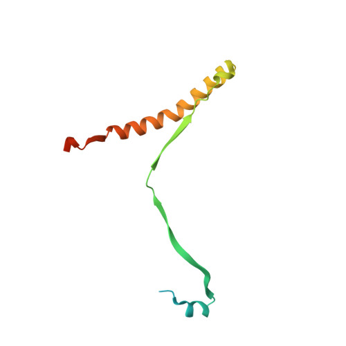
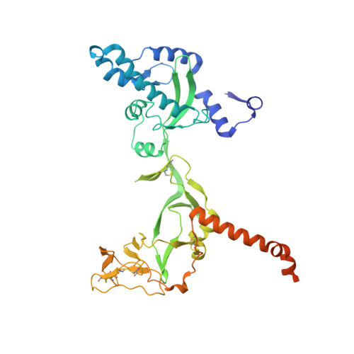
8UUP

Structure of the Measles virus Fusion protein in the pre-fusion conformation


Experimental Data Snapshot

  • Method: ELECTRON MICROSCOPY
  • Resolution: 2.11 Å
  • Aggregation State: PARTICLE 
  • Reconstruction Method: SINGLE PARTICLE 

Starting Model: experimental
View more details

wwPDB Validation   3D Report Full Report


This is version 1.3 of the entry. See complete history


Literature

A neutralizing antibody prevents postfusion transition of measles virus fusion protein.

Zyla, D.S.Della Marca, R.Niemeyer, G.Zipursky, G.Stearns, K.Leedale, C.Sobolik, E.B.Callaway, H.M.Hariharan, C.Peng, W.Parekh, D.Marcink, T.C.Diaz Avalos, R.Horvat, B.Mathieu, C.Snijder, J.Greninger, A.L.Hastie, K.M.Niewiesk, S.Moscona, A.Porotto, M.Ollmann Saphire, E.

(2024) Science 384: eadm8693-eadm8693

  • DOI: https://doi.org/10.1126/science.adm8693
  • Primary Citation of Related Structures:  
    8UT2, 8UTF, 8UUP, 8UUQ, 9AT8

  • PubMed Abstract: 

    Measles virus (MeV) presents a public health threat that is escalating as vaccine coverage in the general population declines and as populations of immunocompromised individuals, who cannot be vaccinated, increase. There are no approved therapeutics for MeV. Neutralizing antibodies targeting viral fusion are one potential therapeutic approach but have not yet been structurally characterized or advanced to clinical use. We present cryo-electron microscopy (cryo-EM) structures of prefusion F alone [2.1-angstrom (Å) resolution], F complexed with a fusion-inhibitory peptide (2.3-Å resolution), F complexed with the neutralizing and protective monoclonal antibody (mAb) 77 (2.6-Å resolution), and an additional structure of postfusion F (2.7-Å resolution). In vitro assays and examination of additional EM classes show that mAb 77 binds prefusion F, arrests F in an intermediate state, and prevents transition to the postfusion conformation. These structures shed light on antibody-mediated neutralization that involves arrest of fusion proteins in an intermediate state.


  • Organizational Affiliation

    Center for Vaccine Innovation, La Jolla Institute for Immunology, La Jolla, CA 92037, USA.


Macromolecules
Find similar proteins by:  (by identity cutoff)  |  3D Structure
Entity ID: 1
MoleculeChains Sequence LengthOrganismDetailsImage
Fusion glycoprotein F0
A, C, E
112Cuspidothrix issatschenkoi B5Mutation(s): 0 
UniProt
Find proteins for Q786F3 (Measles virus (strain Ichinose-B95a))
Explore Q786F3 
Go to UniProtKB:  Q786F3
Entity Groups  
Sequence Clusters30% Identity50% Identity70% Identity90% Identity95% Identity100% Identity
UniProt GroupQ786F3
Glycosylation
Glycosylation Sites: 3
Sequence Annotations
Expand
  • Reference Sequence
Find similar proteins by:  (by identity cutoff)  |  3D Structure
Entity ID: 2
MoleculeChains Sequence LengthOrganismDetailsImage
Fusion glycoprotein F0
B, D, F
420Measles virus strain Ichinose-B95aMutation(s): 2 
UniProt
Find proteins for Q786F3 (Measles virus (strain Ichinose-B95a))
Explore Q786F3 
Go to UniProtKB:  Q786F3
Entity Groups  
Sequence Clusters30% Identity50% Identity70% Identity90% Identity95% Identity100% Identity
UniProt GroupQ786F3
Sequence Annotations
Expand
  • Reference Sequence
Oligosaccharides

Help

Entity ID: 3
MoleculeChains Length2D Diagram Glycosylation3D Interactions
beta-D-mannopyranose-(1-4)-2-acetamido-2-deoxy-beta-D-glucopyranose-(1-4)-2-acetamido-2-deoxy-beta-D-glucopyranose
G
3N-Glycosylation
Glycosylation Resources
GlyTouCan:  G15407YE
GlyCosmos:  G15407YE
GlyGen:  G15407YE
Entity ID: 4
MoleculeChains Length2D Diagram Glycosylation3D Interactions
2-acetamido-2-deoxy-beta-D-glucopyranose-(1-4)-2-acetamido-2-deoxy-beta-D-glucopyranose
H, J, L
2N-Glycosylation
Glycosylation Resources
GlyTouCan:  G42666HT
GlyCosmos:  G42666HT
GlyGen:  G42666HT
Entity ID: 5
MoleculeChains Length2D Diagram Glycosylation3D Interactions
alpha-D-mannopyranose-(1-3)-[alpha-D-mannopyranose-(1-6)]beta-D-mannopyranose-(1-4)-2-acetamido-2-deoxy-beta-D-glucopyranose-(1-4)-2-acetamido-2-deoxy-beta-D-glucopyranose
I, K
5N-Glycosylation
Glycosylation Resources
GlyTouCan:  G22768VO
GlyCosmos:  G22768VO
GlyGen:  G22768VO
Experimental Data & Validation

Experimental Data

  • Method: ELECTRON MICROSCOPY
  • Resolution: 2.11 Å
  • Aggregation State: PARTICLE 
  • Reconstruction Method: SINGLE PARTICLE 
EM Software:
TaskSoftware PackageVersion
RECONSTRUCTIONcryoSPARCv4.3
MODEL REFINEMENTPHENIX

Structure Validation

View Full Validation Report


Entry History & Funding Information

Deposition Data


Funding OrganizationLocationGrant Number
Swiss National Science FoundationSwitzerlandP2EZP3_195680
Swiss National Science FoundationSwitzerlandP500PB_210992
National Institutes of Health/National Institute of Neurological Disorders and Stroke (NIH/NINDS)United StatesNS105699
National Institutes of Health/National Institute of Neurological Disorders and Stroke (NIH/NINDS)United StatesNS091263
National Institutes of Health/National Institute Of Allergy and Infectious Diseases (NIH/NIAID)United StatesAI176833

Revision History  (Full details and data files)

  • Version 1.0: 2024-07-03
    Type: Initial release
  • Version 1.1: 2024-07-10
    Changes: Data collection, Database references
  • Version 1.2: 2024-07-24
    Changes: Data collection, Source and taxonomy
  • Version 1.3: 2024-11-13
    Changes: Data collection, Structure summary