A "terminal" case of glycan catabolism: Structural and enzymatic characterization of the sialidases of Clostridium perfringens.
Medley, B.J., Low, K.E., Irungu, J.D.W., Kipchumba, L., Daneshgar, P., Liu, L., Garber, J.M., Klassen, L., Inglis, G.D., Boons, G.J., Zandberg, W.F., Abbott, D.W., Boraston, A.B.(2024) J Biol Chem 300: 107750-107750
- PubMed: 39251137 Search on PubMed
- DOI: https://doi.org/10.1016/j.jbc.2024.107750
- Primary Citation Related Structures:
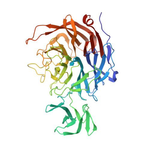
8U2A, 8U5O, 8UB5, 8UL7, 8ULE, 8UM0, 8URL, 8UVV, 9C20 - PubMed Abstract:
Sialic acids are commonly found on the terminal ends of biologically important carbohydrates, including intestinal mucin O-linked glycans. Pathogens such as Clostridium perfringens, the causative agent of necrotic enteritis (NE) in poultry and humans, have the ability to degrade host mucins and colonize the mucus layer, which involves removal of the terminal sialic acid by carbohydrate-active enzymes (CAZymes). Here we present the structural and biochemical characterization of the GH33 catalytic domains of the three sialidases of C. perfringens and probe their substrate specificity. The catalytically active domains, which we refer to as NanH GH33 , NanJ GH33 , and NanI GH33 , displayed differential activity on various naturally occurring forms of sialic acid. We report the X-ray crystal structures of these domains in complex with relevant sialic acid variants revealing the molecular basis of how each catalytic domain accommodates different sialic acids. NanH GH33 displays a distinct preference for α-2,3-linked sialic acid, but can process α-2,6-linked sialic acid. NanJ GH33 and NanI GH33 both exhibit the ability to process α-2,3- and α-2,6-linked sialic acid without any significant apparent preference. All three enzymes were sensitive to generic and commercially available sialidase inhibitors, which impeded sialidase activity in cultures as well as the growth of C. perfringens on sialylated glycans. The knowledge gained in these studies can be applied to in vivo models for C. perfringens growth and metabolism of mucin O-glycans, with a view towards future mitigation of bacterial colonization and infection of intestinal tissues.
- Department of Biochemistry & Microbiology, University of Victoria, Victoria, BC, V8P 5C2 Canada.
Organizational Affiliation: