Disrupting the network of co-evolving amino terminal domain residues relieves mitochondrial calcium uptake inhibition by MCUb.
Colussi, D.M., Grainger, R., Noble, M., Lake, T., Junop, M., Stathopulos, P.B.(2025) Comput Struct Biotechnol J 27: 190-213
- PubMed: 40017731 Search on PubMedSearch on PubMed Central
- DOI: https://doi.org/10.1016/j.csbj.2024.12.007
- Primary Citation Related Structures:
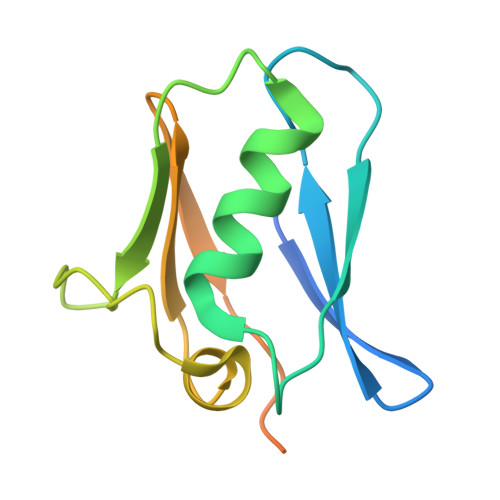
8URG - PubMed Abstract:
The regulatory mechanisms of the mitochondrial calcium uniporter complex (mtCU), the predominant channel mediating calcium (Ca 2 + ) flux into the matrix, are critical for bioenergetics and cell fate. The pore-forming components of mtCU are the mitochondrial Ca 2+ uniporter (MCU) subunit and the MCU dominant-negative beta (MCUb) subunit. Despite both MCU paralogs having conserved Asp-Ile-Met-Glu motifs responsible for Ca 2+ selectivity, MCUb mediates only low Ca 2+ conduction and has been characterized as an inhibitory subunit. We previously identified the MCU amino-terminal domain (NTD) as a negative feedback regulator of mtCU upon divalent cation binding but the role of the MCUb-NTD remains unknown. Thus, to gain mechanistic insight into the competing MCU and MCUb functions, we here studied the divalent cation binding properties of the MCU- and MCUb-NTDs that tightly interact within and between tetrameric channels. First, we resolved a high-resolution MCU-NTD crystal structure in the absence of divalent ions at 1.6 Å, using this structure to model the homologous MCUb-NTD. Further, we conducted 1 μs all-atom molecular dynamics (MD) simulations in the presence and absence of Ca 2+ and Mg 2+ ions, not only finding increased MCU-NTD stability at high temperatures compared to MCUb-NTD but also discrete Ca 2+ -binding sites on the two domains. Remarkably, the distinct Ca 2+ binding site on the central α-helix of MCUb-NTD was also identified in a functional sector of co-evolving residues, with either direct mutation to the coordinating residues or mutation to a separate site within the sector disrupting Ca 2+ binding in silico and in vitro as well as enhancing mitochondrial Ca 2+ uptake in cellulo . Thus, we reveal that matrix Ca 2+ binding to both the MCU-NTD and MCUb-NTD promote mtCU inhibition through disparate interaction sites, highlighting the evolution of discrete feedback regulation mechanisms to precisely control mtCU function.
- Department of Physiology and Pharmacology, Schulich School of Medicine and Dentistry, University of Western Ontario, N6A5C1, Canada.
Organizational Affiliation: