8UR9 | pdb_00008ur9

Crystal Structure of the SARS-CoV-2 Main Protease in Complex with Compound 61


Experimental Data Snapshot

  • Method: X-RAY DIFFRACTION
  • Resolution: 2.30 Å
  • R-Value Free: 
    0.236 (Depositor), 0.235 (DCC) 
  • R-Value Work: 
    0.203 (Depositor), 0.204 (DCC) 
  • R-Value Observed: 
    0.205 (Depositor) 

Starting Model: experimental
View more details

wwPDB Validation 3D Report Full Report

Validation slider image for 8UR9

Ligand Structure Quality Assessment 


This is version 1.0 of the entry. See complete history

Literature

Proof-of-concept studies with a computationally designed M pro inhibitor as a synergistic combination regimen alternative to Paxlovid.

Papini, C.Ullah, I.Ranjan, A.P.Zhang, S.Wu, Q.Spasov, K.A.Zhang, C.Mothes, W.Crawford, J.M.Lindenbach, B.D.Uchil, P.D.Kumar, P.Jorgensen, W.L.Anderson, K.S.

(2024) Proc Natl Acad Sci U S A 121: e2320713121-e2320713121

  • DOI: https://doi.org/10.1073/pnas.2320713121
  • Primary Citation Related Structures: 
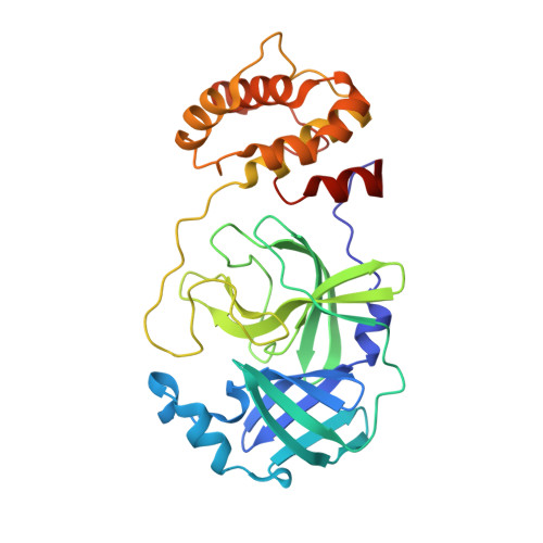
    8UR9

  • PubMed Abstract: 

    As the SARS-CoV-2 virus continues to spread and mutate, it remains important to focus not only on preventing spread through vaccination but also on treating infection with direct-acting antivirals (DAA). The approval of Paxlovid, a SARS-CoV-2 main protease (M pro ) DAA, has been significant for treatment of patients. A limitation of this DAA, however, is that the antiviral component, nirmatrelvir, is rapidly metabolized and requires inclusion of a CYP450 3A4 metabolic inhibitor, ritonavir, to boost levels of the active drug. Serious drug-drug interactions can occur with Paxlovid for patients who are also taking other medications metabolized by CYP4503A4, particularly transplant or otherwise immunocompromised patients who are most at risk for SARS-CoV-2 infection and the development of severe symptoms. Developing an alternative antiviral with improved pharmacological properties is critical for treatment of these patients. By using a computational and structure-guided approach, we were able to optimize a 100 to 250 μM screening hit to a potent nanomolar inhibitor and lead compound, Mpro61. In this study, we further evaluate Mpro61 as a lead compound, starting with examination of its mode of binding to SARS-CoV-2 M pro . In vitro pharmacological profiling established a lack of off-target effects, particularly CYP450 3A4 inhibition, as well as potential for synergy with the currently approved alternate antiviral, molnupiravir. Development and subsequent testing of a capsule formulation for oral dosing of Mpro61 in B6-K18-hACE2 mice demonstrated favorable pharmacological properties, efficacy, and synergy with molnupiravir, and complete recovery from subsequent challenge by SARS-CoV-2, establishing Mpro61 as a promising potential preclinical candidate.


  • Organizational Affiliation
    • Department of Pharmacology, Yale University School of Medicine, New Haven, CT 06520-8066.

Macromolecule Content 

  • Total Structure Weight: 68.22 kDa 
  • Atom Count: 4,497 
  • Modeled Residue Count: 594 
  • Deposited Residue Count: 612 
  • Unique protein chains: 1

Macromolecules

Find similar proteins by:|  3D Structure
Entity ID: 1
MoleculeChains  Sequence LengthOrganismDetailsImage
3C-like proteinase nsp5
A, B
306Severe acute respiratory syndrome coronavirus 2Mutation(s): 0 
Gene Names: rep1a-1b
EC: 3.4.22.69
UniProt
Find proteins for P0DTD1 (Severe acute respiratory syndrome coronavirus 2)
Explore P0DTD1 
Go to UniProtKB:  P0DTD1
Entity Groups
Sequence Clusters30% Identity50% Identity70% Identity90% Identity95% Identity100% Identity
UniProt GroupP0DTD1
Sequence Annotations
Expand
Reference Sequence

Small Molecules

Ligands 1 Unique
IDChains Name / Formula / InChI Key2D Diagram3D Interactions
XEK
(Subject of Investigation/LOI)

Query on XEK


Download:Ideal Coordinates CCD File
C [auth A](5P)-5-[(1P,3M,3'P)-3-{3-chloro-5-[(2-chlorophenyl)methoxy]-4-fluorophenyl}-2-oxo-2H-[1,3'-bipyridin]-5-yl]-1-methylpyrimidine-2,4(1H,3H)-dione
C28 H19 Cl2 F N4 O4
KIJKUSPNZALBLD-UHFFFAOYSA-N

Experimental Data & Validation

Experimental Data

  • Method: X-RAY DIFFRACTION
  • Resolution: 2.30 Å
  • R-Value Free:  0.236 (Depositor), 0.235 (DCC) 
  • R-Value Work:  0.203 (Depositor), 0.204 (DCC) 
  • R-Value Observed: 0.205 (Depositor) 
Space Group: P 21 21 21
Unit Cell:
Length ( Å )Angle ( ˚ )
a = 67.91α = 90
b = 99.895β = 90
c = 104.314γ = 90
Software Package:
Software NamePurpose
PHENIXrefinement
XDSdata reduction
XDSdata scaling
PHENIXphasing

Structure Validation

View Full Validation Report


Ligand Structure Quality Assessment 


Entry History 

& Funding Information

Deposition Data


Funding OrganizationLocationGrant Number
National Institutes of Health/National Institute of General Medical Sciences (NIH/NIGMS)United StatesP30 GM124165
Other governmentUnited StatesNIH-ORIP HEI grant (S10OD021527)
Department of Energy (DOE, United States)United StatesDE-AC02-06CH11357

Revision History  (Full details and data files)

  • Version 1.0: 2024-05-15
    Type: Initial release