


Streptococcus surface alpha enolase exposed dimers were found to be the active form on lipid surface that binds to human plasminogen
Tjia-Fleck, S., Readnour, B.M., Castellino, F.J.To be published.
Experimental Data Snapshot
wwPDB Validation 3D Report Full Report

Entity ID: 1 | |||||
|---|---|---|---|---|---|
| Molecule | Chains | Sequence Length | Organism | Details | Image |
| Plasminogen | 791 | Homo sapiens | Mutation(s): 0 Gene Names: PLG | 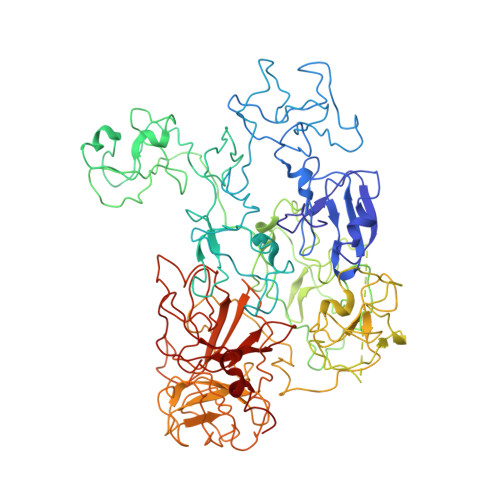 | |
UniProt & NIH Common Fund Data Resources | |||||
PHAROS: P00747 GTEx: ENSG00000122194 | |||||
Entity Groups | |||||
| Sequence Clusters | 30% Identity50% Identity70% Identity90% Identity95% Identity100% Identity | ||||
| UniProt Group | P00747 | ||||
Sequence AnnotationsExpand | |||||
Reference Sequence | |||||
| Funding Organization | Location | Grant Number |
|---|---|---|
| National Institutes of Health/National Heart, Lung, and Blood Institute (NIH/NHLBI) | United States | HL013423 |


