


Mapping of the Reaction Trajectory catalyzed by Class I Ketol-Acid Reductoisomerase
Lin, X., Lonhienne, T., Lv, Y., Kurz, J., McGeary, R., Schenk, G., Guddat, L.W.(2024) ACS Catal
Experimental Data Snapshot
Starting Model: experimental
View more details
(2024) ACS Catal
Entity ID: 1 | |||||
|---|---|---|---|---|---|
| Molecule | Chains | Sequence Length | Organism | Details | Image |
| Ketol-acid reductoisomerase | 330 | Campylobacter jejuni | Mutation(s): 0 Gene Names: ilvC, CW563_00670 EC: 1.1.1.86 | 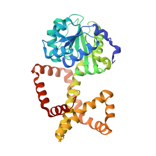 | |
UniProt | |||||
Entity Groups | |||||
| Sequence Clusters | 30% Identity50% Identity70% Identity90% Identity95% Identity100% Identity | ||||
| UniProt Group | Q9PHN5 | ||||
Sequence AnnotationsExpand | |||||
Reference Sequence | |||||
| Ligands 3 Unique | |||||
|---|---|---|---|---|---|
| ID | Chains | Name / Formula / InChI Key | 2D Diagram | 3D Interactions | |
| NDP (Subject of Investigation/LOI) Download:Ideal Coordinates CCD File | BA [auth I] DB [auth F] EA [auth D] HB [auth A] IA [auth G] | NADPH DIHYDRO-NICOTINAMIDE-ADENINE-DINUCLEOTIDE PHOSPHATE C21 H30 N7 O17 P3 ACFIXJIJDZMPPO-NNYOXOHSSA-N |  | ||
| X9W (Subject of Investigation/LOI) Download:Ideal Coordinates CCD File | AA [auth I] AB [auth F] EB [auth A] FA [auth D] JA [auth G] | (2R)-(dimethylphosphoryl)(hydroxy)acetic acid C4 H9 O4 P UTDPHALOLFEIHB-SCSAIBSYSA-N |  | ||
| MG Download:Ideal Coordinates CCD File | BB [auth F] CA [auth D] CB [auth F] DA [auth D] FB [auth A] | MAGNESIUM ION Mg JLVVSXFLKOJNIY-UHFFFAOYSA-N |  | ||
| Length ( Å ) | Angle ( ˚ ) |
|---|---|
| a = 111.947 | α = 110.91 |
| b = 111.954 | β = 107.73 |
| c = 111.944 | γ = 109.76 |
| Software Name | Purpose |
|---|---|
| PHENIX | refinement |
| XDS | data reduction |
| XDS | data scaling |
| PHENIX | phasing |
| Funding Organization | Location | Grant Number |
|---|---|---|
| Australian Research Council (ARC) | Australia | DP210101802 |


