8UM6 | pdb_00008um6

Structure of copper bound to YcnI W137F


Experimental Data Snapshot

  • Method: X-RAY DIFFRACTION
  • Resolution: 1.95 Å
  • R-Value Free: 
    0.239 (Depositor), 0.242 (DCC) 
  • R-Value Work: 
    0.203 (Depositor), 0.208 (DCC) 
  • R-Value Observed: 
    0.210 (Depositor) 

Starting Model: experimental
View more details

wwPDB Validation 3D Report Full Report

Validation slider image for 8UM6

This is version 1.1 of the entry. See complete history

Literature

Stabilization of a Cu-binding site by a highly conserved tryptophan residue.

de Oliveira Silva, Y.R.Zheng, D.Peters, S.C.Fisher, O.S.

(2024) J Inorg Biochem 253: 112501-112501

  • DOI: https://doi.org/10.1016/j.jinorgbio.2024.112501
  • Primary Citation Related Structures: 
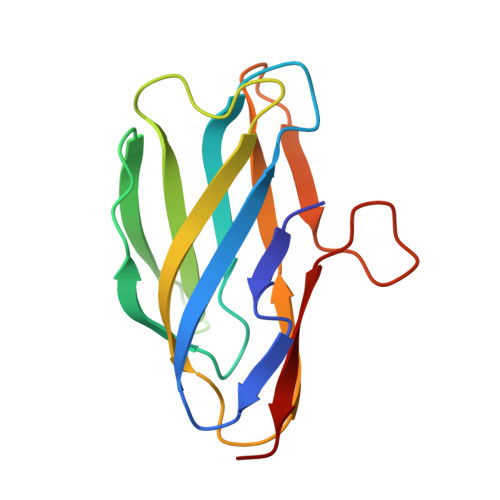
    8UM6

  • PubMed Abstract: 

    Copper serves as an essential cofactor for nearly all living organisms. There are still many gaps remaining in our knowledge of how Gram-positive bacteria import copper and maintain homeostasis. To obtain a better understanding of how these processes work, here we focus on the ycnKJI operon responsible for regulating copper levels in the Gram-positive bacterium Bacillus subtilis. This operon encodes three Cu-related proteins: a copper-dependent transcriptional repressor (YcnK), a putative copper importer (YcnJ), and a copper-binding protein of unknown function (YcnI). We previously found that YcnI's extracellular Domain of Unknown Function 1775 (DUF1775) houses a monohistidine brace motif that coordinates a single Cu(II) ion. The Cu(II) binding site includes a highly conserved tryptophan residue. Here, we investigate the role of that tryptophan and the ability of the protein to interact with other oxidation states of Cu. We find that YcnI exhibits strong preference for binding Cu in the oxidized Cu(II) state, and that the conserved tryptophan residue is not essential for the interaction. We determine the structure of a tryptophan variant to 1.95 Å resolution that indicates that the tryptophan is needed to stabilize the metal binding interaction, and find that this variant has weaker affinity for Cu(II) than the wild-type protein. Together, these data provide further insights into the DUF1775 domain family and reveal the role of the conserved tryptophan residue.


  • Organizational Affiliation
    • Department of Chemistry, Lehigh University, 6 E Packer Ave, Bethlehem, PA 18015, USA.

Macromolecule Content 

  • Total Structure Weight: 28.94 kDa 
  • Atom Count: 2,200 
  • Modeled Residue Count: 257 
  • Deposited Residue Count: 258 
  • Unique protein chains: 1

Macromolecules

Find similar proteins by:|  3D Structure
Entity ID: 1
MoleculeChains  Sequence LengthOrganismDetailsImage
Uncharacterized protein YcnI
A, B
129Bacillus subtilisMutation(s): 1 
Gene Names: ycnI
UniProt
Find proteins for P94431 (Bacillus subtilis (strain 168))
Explore P94431 
Go to UniProtKB:  P94431
Entity Groups
Sequence Clusters30% Identity50% Identity70% Identity90% Identity95% Identity100% Identity
UniProt GroupP94431
Sequence Annotations
Expand
Reference Sequence

Experimental Data & Validation

Experimental Data

  • Method: X-RAY DIFFRACTION
  • Resolution: 1.95 Å
  • R-Value Free:  0.239 (Depositor), 0.242 (DCC) 
  • R-Value Work:  0.203 (Depositor), 0.208 (DCC) 
  • R-Value Observed: 0.210 (Depositor) 
Space Group: P 63 2 2
Unit Cell:
Length ( Å )Angle ( ˚ )
a = 90.263α = 90
b = 90.263β = 90
c = 207.508γ = 120
Software Package:
Software NamePurpose
PHENIXrefinement
DIALSdata reduction
DIALSdata scaling
PHASERphasing

Structure Validation

View Full Validation Report


Entry History 

& Funding Information

Deposition Data


Funding OrganizationLocationGrant Number
National Science Foundation (NSF, United States)United States2137107

Revision History  (Full details and data files)

  • Version 1.0: 2024-02-07
    Type: Initial release
  • Version 1.1: 2024-02-21
    Changes: Database references