Structural mechanism of proton conduction in otopetrin proton channel.
Gan, N., Zeng, W., Han, Y., Chen, Q., Jiang, Y.(2024) Nat Commun 15: 7250-7250
- PubMed: 39179582 Search on PubMedSearch on PubMed Central
- DOI: https://doi.org/10.1038/s41467-024-51803-x
- Primary Citation Related Structures:
8UG4, 8UG5, 8UG6, 8UG7, 8UG8, 8UGA - PubMed Abstract:
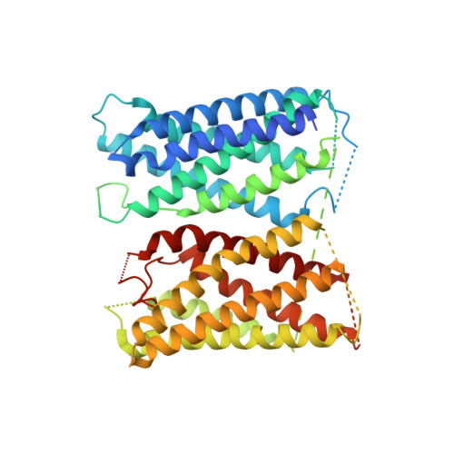
The otopetrin (OTOP) proteins were recently characterized as extracellular proton-activated proton channels. Several recent OTOP channel structures demonstrated that the channels form a dimer with each subunit adopting a double-barrel architecture. However, the structural mechanisms underlying some basic functional properties of the OTOP channels remain unresolved, including extracellular pH activation, proton conducting pathway, and rapid desensitization. In this study, we performed structural and functional characterization of the Caenorhabditis elegans OTOP8 (CeOTOP8) and mouse OTOP2 (mOTOP2) and illuminated a set of conformational changes related to the proton-conducting process in OTOP. The structures of CeOTOP8 reveal the conformational change at the N-terminal part of TM12 that renders the channel in a transiently proton-transferring state, elucidating an inter-barrel, Glu/His-bridged proton passage within each subunit. The structures of mOTOP2 reveal the conformational change at the N-terminal part of TM6 that exposes the central glutamate to the extracellular solution for protonation. In addition, the structural comparison between CeOTOP8 and mOTOP2, along with the structure-based mutagenesis, demonstrates that an inter-subunit movement at the OTOP channel dimer interface plays a central role in regulating channel activity. Combining the structural information from both channels, we propose a working model describing the multi-step conformational changes during the proton conducting process.
- Howard Hughes Medical Institute and Department of Physiology, University of Texas Southwestern Medical Center, Dallas, TX, USA.
Organizational Affiliation: