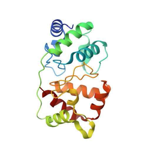
The structure of the diheme cytochrome c 4 from Neisseria gonorrhoeae reveals multiple contributors to tuning reduction potentials.
Zhong, F., Reik, M.E., Ragusa, M.J., Pletneva, E.V.(2024) J Inorg Biochem 253: 112496-112496
- PubMed: 38330683
- DOI: https://doi.org/10.1016/j.jinorgbio.2024.112496
- Primary Citation of Related Structures:
8UF3 - PubMed Abstract:
Cytochrome c 4 (c 4 ) is a diheme protein implicated as an electron donor to cbb 3 oxidases in multiple pathogenic bacteria. Despite its prevalence, understanding of how specific structural features of c 4 optimize its function is lacking. The human pathogen Neisseria gonorrhoeae (Ng) thrives in low oxygen environments owing to the activity of its cbb 3 oxidase. Herein, we report characterization of Ng c 4 . Spectroelectrochemistry experiments of the wild-type (WT) protein have shown that the two Met/His-ligated hemes differ in potentials by ∼100 mV, and studies of the two His/His-ligated variants provided unambiguous assignment of heme A from the N-terminal domain of the protein as the high-potential heme. The crystal structure of the WT protein at 2.45 Å resolution has revealed that the two hemes differ in their solvent accessibility. In particular, interactions made by residues His57 and Ser59 in Loop1 near the axial ligand Met63 contribute to the tight enclosure of heme A, working together with the surface charge, to raise the reduction potential of the heme iron in this domain. The structure reveals a prominent positively-charged patch, which encompasses surfaces of both domains. In contrast to prior findings with c 4 from Pseudomonas stutzeri, the interdomain interface of Ng c 4 contributes minimally to the values of the heme iron potentials in the two domains. Analyses of the heme solvent accessibility, interface properties, and surface charges offer insights into the interplay of these structural elements in tuning redox properties of c 4 and other multiheme proteins.
- Department of Chemistry, Dartmouth College, Hanover, NH 03755, United States.
Organizational Affiliation:


