8UEL

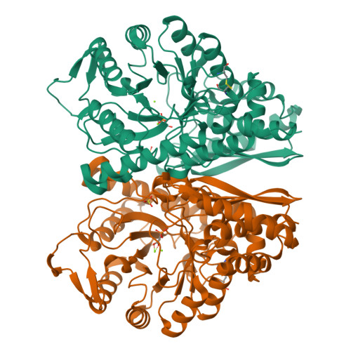
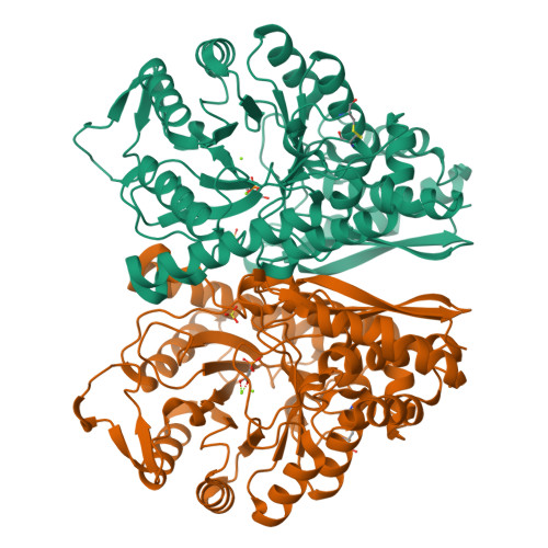
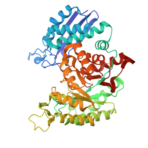
Crystal structure of enolase from Litopenaeus vannamei

  • Classification: LYASE
  • Organism(s): Penaeus vannamei
  • Mutation(s): No 

  • Deposited: 2023-10-01 Released: 2023-12-13 
  • Deposition Author(s): Chang, X., Zhao, G.
  • Funding Organization(s): Other government

Experimental Data Snapshot

  • Method: X-RAY DIFFRACTION
  • Resolution: 2.49 Å
  • R-Value Free: 0.224 
  • R-Value Work: 0.168 
  • R-Value Observed: 0.171 

Starting Model: experimental
View more details

wwPDB Validation   3D Report Full Report


Ligand Structure Quality Assessment 


This is version 1.2 of the entry. See complete history


Literature

Characterization and Structural Analyses of Enolase from Shrimp ( Litopenaeus vannamei ).

Chang, X.Zhang, T.Zang, J.Lv, C.Zhao, G.

(2023) J Agric Food Chem 71: 19783-19790

  • DOI: https://doi.org/10.1021/acs.jafc.3c07135
  • Primary Citation of Related Structures:  
    8UEL

  • PubMed Abstract: 

    Transcriptome analysis had recognized enolase from shrimp Litopenaeus vannamei ( L. vannamei ), which is termed LvEnolase, as one of the allergens, but its amino acid sequence and protein structure have been lacking. In this study, natural LvEnolase was isolated from L. vannamei and characterized for the first time. The full-length cDNA sequence of LvEnolase was effectively cloned, which encoded 434 amino acid residues. The crystal structure of LvEnolase was successfully determined at a resolution of 2.5 Å by X-ray crystallography (PDB: 8UEL). Notably, it was observed that near the active center, a loop exists in either an open or closed state, and the open loop was associated with the product release phase. Furthermore, enzyme activity assays were conducted to validate the catalytic capabilities of purified LvEnolase. These findings significantly enhance our comprehension of the enolase family and provide valuable support for delving into the functions and characteristics of LvEnolase.


  • Organizational Affiliation

    College of Food Science & Nutritional Engineering, China Agricultural University, Beijing 100083, China.


Macromolecules
Find similar proteins by:  (by identity cutoff)  |  3D Structure
Entity ID: 1
MoleculeChains Sequence LengthOrganismDetailsImage
Enolase
A, B
433Penaeus vannameiMutation(s): 0 
EC: 4.2.1.11
UniProt
Find proteins for O96656 (Penaeus monodon)
Explore O96656 
Go to UniProtKB:  O96656
Entity Groups  
Sequence Clusters30% Identity50% Identity70% Identity90% Identity95% Identity100% Identity
UniProt GroupO96656
Sequence Annotations
Expand
  • Reference Sequence
Small Molecules
Ligands 3 Unique
IDChains Name / Formula / InChI Key2D Diagram3D Interactions
PEP (Subject of Investigation/LOI)
Query on PEP

Download Ideal Coordinates CCD File 
C [auth A],
G [auth B]
PHOSPHOENOLPYRUVATE
C3 H5 O6 P
DTBNBXWJWCWCIK-UHFFFAOYSA-N
SO4
Query on SO4

Download Ideal Coordinates CCD File 
F [auth A],
J [auth B]
SULFATE ION
O4 S
QAOWNCQODCNURD-UHFFFAOYSA-L
MG (Subject of Investigation/LOI)
Query on MG

Download Ideal Coordinates CCD File 
D [auth A],
E [auth A],
H [auth B],
I [auth B]
MAGNESIUM ION
Mg
JLVVSXFLKOJNIY-UHFFFAOYSA-N
Experimental Data & Validation

Experimental Data

  • Method: X-RAY DIFFRACTION
  • Resolution: 2.49 Å
  • R-Value Free: 0.224 
  • R-Value Work: 0.168 
  • R-Value Observed: 0.171 
  • Space Group: P 21 21 21
Unit Cell:
Length ( Å )Angle ( ˚ )
a = 79.613α = 90
b = 86.571β = 90
c = 155.881γ = 90
Software Package:
Software NamePurpose
PHENIXrefinement
HKL-3000data scaling
HKL-3000data reduction
MOLREPphasing

Structure Validation

View Full Validation Report


Ligand Structure Quality Assessment 


Entry History & Funding Information

Deposition Data


Funding OrganizationLocationGrant Number
Other governmentChina2021YDF2100105

Revision History  (Full details and data files)

  • Version 1.0: 2023-12-13
    Type: Initial release
  • Version 1.1: 2023-12-27
    Changes: Database references
  • Version 1.2: 2024-11-20
    Changes: Structure summary