8UEE

Atomic structure of Salmonella SipA/F-actin complex by cryo-EM


Experimental Data Snapshot

  • Method: ELECTRON MICROSCOPY
  • Resolution: 3.20 Å
  • Aggregation State: FILAMENT 
  • Reconstruction Method: HELICAL 

wwPDB Validation   3D Report Full Report


This is version 1.1 of the entry. See complete history


Literature

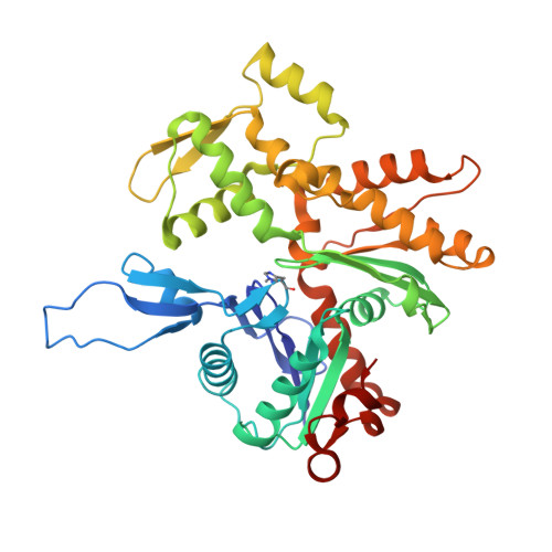
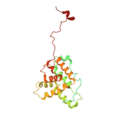
Stabilization of F-actin by Salmonella effector SipA resembles the structural effects of inorganic phosphate and phalloidin.

Niedzialkowska, E.Runyan, L.A.Kudryashova, E.Egelman, E.H.Kudryashov, D.S.

(2024) Structure 32: 725-738.e8

  • DOI: https://doi.org/10.1016/j.str.2024.02.022
  • Primary Citation of Related Structures:  
    8UEE

  • PubMed Abstract: 

    Entry of Salmonella into host enterocytes relies on its pathogenicity island 1 effector SipA. We found that SipA binds to F-actin in a 1:2 stoichiometry with sub-nanomolar affinity. A cryo-EM reconstruction revealed that SipA's globular core binds at the groove between actin strands, whereas the extended C-terminal arm penetrates deeply into the inter-strand space, stabilizing F-actin from within. The unusually strong binding of SipA is achieved by a combination of fast association via the core and very slow dissociation dictated by the arm. Similar to P i , BeF 3 , and phalloidin, SipA potently inhibited actin depolymerization by actin depolymerizing factor (ADF)/cofilin, which correlated with increased filament stiffness, supporting the hypothesis that F-actin's mechanical properties contribute to the recognition of its nucleotide state by protein partners. The remarkably strong binding to F-actin maximizes the toxin's effects at the injection site while minimizing global influence on the cytoskeleton and preventing pathogen detection by the host cell.


  • Organizational Affiliation

    Department of Biochemistry and Molecular Genetics, University of Virginia, Charlottesville, VA 22903, USA.


Macromolecules
Find similar proteins by:  (by identity cutoff)  |  3D Structure
Entity ID: 1
MoleculeChains Sequence LengthOrganismDetailsImage
Actin, alpha skeletal muscle375Oryctolagus cuniculusMutation(s): 0 
UniProt
Find proteins for P68135 (Oryctolagus cuniculus)
Explore P68135 
Go to UniProtKB:  P68135
Entity Groups  
Sequence Clusters30% Identity50% Identity70% Identity90% Identity95% Identity100% Identity
UniProt GroupP68135
Sequence Annotations
Expand
  • Reference Sequence
Find similar proteins by:  (by identity cutoff)  |  3D Structure
Entity ID: 2
MoleculeChains Sequence LengthOrganismDetailsImage
Cell invasion protein SipAH [auth B],
I [auth C],
J [auth D],
K [auth A]
261Salmonella enterica subsp. enterica serovar Typhimurium str. LT2Mutation(s): 0 
Gene Names: sipAsspASTM2882
UniProt
Find proteins for P0CL52 (Salmonella typhimurium (strain LT2 / SGSC1412 / ATCC 700720))
Explore P0CL52 
Go to UniProtKB:  P0CL52
Entity Groups  
Sequence Clusters30% Identity50% Identity70% Identity90% Identity95% Identity100% Identity
UniProt GroupP0CL52
Sequence Annotations
Expand
  • Reference Sequence
Small Molecules
Ligands 3 Unique
IDChains Name / Formula / InChI Key2D Diagram3D Interactions
ADP (Subject of Investigation/LOI)
Query on ADP

Download Ideal Coordinates CCD File 
AA [auth L]
DA [auth M]
L [auth F]
O [auth H]
R [auth I]
AA [auth L],
DA [auth M],
L [auth F],
O [auth H],
R [auth I],
U [auth J],
X [auth K]
ADENOSINE-5'-DIPHOSPHATE
C10 H15 N5 O10 P2
XTWYTFMLZFPYCI-KQYNXXCUSA-N
PO4 (Subject of Investigation/LOI)
Query on PO4

Download Ideal Coordinates CCD File 
CA [auth L]
FA [auth M]
N [auth F]
Q [auth H]
T [auth I]
CA [auth L],
FA [auth M],
N [auth F],
Q [auth H],
T [auth I],
W [auth J],
Z [auth K]
PHOSPHATE ION
O4 P
NBIIXXVUZAFLBC-UHFFFAOYSA-K
MG (Subject of Investigation/LOI)
Query on MG

Download Ideal Coordinates CCD File 
BA [auth L]
EA [auth M]
M [auth F]
P [auth H]
S [auth I]
BA [auth L],
EA [auth M],
M [auth F],
P [auth H],
S [auth I],
V [auth J],
Y [auth K]
MAGNESIUM ION
Mg
JLVVSXFLKOJNIY-UHFFFAOYSA-N
Modified Residues  1 Unique
IDChains TypeFormula2D DiagramParent
HIC
Query on HIC
A [auth F]
B [auth H]
C [auth I]
D [auth J]
E [auth K]
A [auth F],
B [auth H],
C [auth I],
D [auth J],
E [auth K],
F [auth L],
G [auth M]
L-PEPTIDE LINKINGC7 H11 N3 O2HIS
Experimental Data & Validation

Experimental Data

  • Method: ELECTRON MICROSCOPY
  • Resolution: 3.20 Å
  • Aggregation State: FILAMENT 
  • Reconstruction Method: HELICAL 
EM Software:
TaskSoftware PackageVersion
MODEL REFINEMENTPHENIX1.3
MODEL REFINEMENTCoot0.9.8.1
RECONSTRUCTIONcryoSPARC4.0.2

Structure Validation

View Full Validation Report


Entry History & Funding Information

Deposition Data


Funding OrganizationLocationGrant Number
National Institutes of Health/National Institute of General Medical Sciences (NIH/NIGMS)United StatesGM122510
National Institutes of Health/National Institute of General Medical Sciences (NIH/NIGMS)United StatesGM114666

Revision History  (Full details and data files)

  • Version 1.0: 2023-12-27
    Type: Initial release
  • Version 1.1: 2024-07-10
    Changes: Data collection, Database references