8UE1

Crystal Structure of Human Fructosamine-3-kinase (FN3K)


Experimental Data Snapshot

  • Method: X-RAY DIFFRACTION
  • Resolution: 2.85 Å
  • R-Value Free: 0.258 
  • R-Value Work: 0.198 

Starting Model: experimental
View more details

wwPDB Validation   3D Report Full Report


Ligand Structure Quality Assessment 


This is version 1.2 of the entry. See complete history


Literature

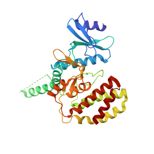
Structural basis for FN3K-mediated protein deglycation.

Lokhandwala, J.Matlack, J.K.Smalley, T.B.Miner 3rd, R.E.Tran, T.H.Binning, J.M.

(2024) Structure 

  • DOI: https://doi.org/10.1016/j.str.2024.07.018
  • Primary Citation of Related Structures:  
    8UE1

  • PubMed Abstract: 

    Protein glycation is a universal, non-enzymatic modification that occurs when a sugar covalently attaches to a primary amine. These spontaneous modifications may have deleterious or regulatory effects on protein function, and their removal is mediated by the conserved metabolic kinase fructosamine-3-kinase (FN3K). Despite its crucial role in protein repair, we currently have a poor understanding of how FN3K engages or phosphorylates its substrates. By integrating structural biology and biochemistry, we elucidated the catalytic mechanism for FN3K-mediated protein deglycation. Our work identifies key amino acids required for binding and phosphorylating glycated substrates and reveals the molecular basis of an evolutionarily conserved protein repair pathway. Additional structural-functional studies revealed unique structural features of human FN3K as well as differences in the dimerization behavior and regulation of FN3K family members. Our findings improve our understanding of the structure of FN3K and its catalytic mechanism, which opens new avenues for therapeutically targeting FN3K.


  • Organizational Affiliation

    Department of Molecular Oncology, H. Lee Moffitt Cancer Center and Research Institute, Tampa, FL 33612, USA.


Macromolecules
Find similar proteins by:  (by identity cutoff)  |  3D Structure
Entity ID: 1
MoleculeChains Sequence LengthOrganismDetailsImage
Fructosamine-3-kinase311Homo sapiensMutation(s): 0 
Gene Names: FN3K
UniProt & NIH Common Fund Data Resources
Find proteins for Q9H479 (Homo sapiens)
Explore Q9H479 
Go to UniProtKB:  Q9H479
PHAROS:  Q9H479
GTEx:  ENSG00000167363 
Entity Groups  
Sequence Clusters30% Identity50% Identity70% Identity90% Identity95% Identity100% Identity
UniProt GroupQ9H479
Sequence Annotations
Expand
  • Reference Sequence
Small Molecules
Ligands 1 Unique
IDChains Name / Formula / InChI Key2D Diagram3D Interactions
EPE (Subject of Investigation/LOI)
Query on EPE

Download Ideal Coordinates CCD File 
B [auth A]4-(2-HYDROXYETHYL)-1-PIPERAZINE ETHANESULFONIC ACID
C8 H18 N2 O4 S
JKMHFZQWWAIEOD-UHFFFAOYSA-N
Experimental Data & Validation

Experimental Data

  • Method: X-RAY DIFFRACTION
  • Resolution: 2.85 Å
  • R-Value Free: 0.258 
  • R-Value Work: 0.198 
  • Space Group: P 1 21 1
Unit Cell:
Length ( Å )Angle ( ˚ )
a = 52.484α = 90
b = 50.241β = 106.33
c = 65.651γ = 90
Software Package:
Software NamePurpose
XSCALEdata scaling
PHENIXrefinement
XDSdata reduction
PHASERphasing

Structure Validation

View Full Validation Report


Ligand Structure Quality Assessment 


Entry History & Funding Information

Deposition Data


Funding OrganizationLocationGrant Number
National Institutes of Health/National Institute of General Medical Sciences (NIH/NIGMS)United States--

Revision History  (Full details and data files)

  • Version 1.0: 2024-08-07
    Type: Initial release
  • Version 1.1: 2024-08-21
    Changes: Structure summary
  • Version 1.2: 2024-09-04
    Changes: Database references