De novo design of pH-responsive self-assembling helical protein filaments.
Shen, H., Lynch, E.M., Akkineni, S., Watson, J.L., Decarreau, J., Bethel, N.P., Benna, I., Sheffler, W., Farrell, D., DiMaio, F., Derivery, E., De Yoreo, J.J., Kollman, J., Baker, D.(2024) Nat Nanotechnol 19: 1016-1021
- PubMed: 38570702 Search on PubMedSearch on PubMed Central
- DOI: https://doi.org/10.1038/s41565-024-01641-1
- Primary Citation Related Structures:
8UAO, 8UB3, 8UBG - PubMed Abstract:
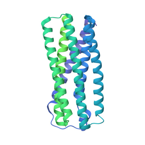
Biological evolution has led to precise and dynamic nanostructures that reconfigure in response to pH and other environmental conditions. However, designing micrometre-scale protein nanostructures that are environmentally responsive remains a challenge. Here we describe the de novo design of pH-responsive protein filaments built from subunits containing six or nine buried histidine residues that assemble into micrometre-scale, well-ordered fibres at neutral pH. The cryogenic electron microscopy structure of an optimized design is nearly identical to the computational design model for both the subunit internal geometry and the subunit packing into the fibre. Electron, fluorescent and atomic force microscopy characterization reveal a sharp and reversible transition from assembled to disassembled fibres over 0.3 pH units, and rapid fibre disassembly in less than 1 s following a drop in pH. The midpoint of the transition can be tuned by modulating buried histidine-containing hydrogen bond networks. Computational protein design thus provides a route to creating unbound nanomaterials that rapidly respond to small pH changes.
- Department of Biochemistry, University of Washington, Seattle, WA, USA. shenh2@uw.edu.
Organizational Affiliation: