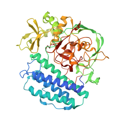
8U41

OvsA from Halomonas utahensis, an ovoselenol-biosynthetic selenoxide synthase in complex with histidine


Experimental Data Snapshot

  • Method: X-RAY DIFFRACTION
  • Resolution: 2.72 Å
  • R-Value Free: 0.246 
  • R-Value Work: 0.225 
  • R-Value Observed: 0.226 

Starting Model: in silico
View more details

wwPDB Validation   3D Report Full Report


Ligand Structure Quality Assessment 


This is version 1.2 of the entry. See complete history


Literature

Discovery of the selenium-containing antioxidant ovoselenol derived from convergent evolution.

Kayrouz, C.M.Ireland, K.A.Ying, V.Y.Davis, K.M.Seyedsayamdost, M.R.

(2024) Nat Chem 16: 1868-1875

  • DOI: https://doi.org/10.1038/s41557-024-01600-2
  • Primary Citation of Related Structures:  
    8U41, 8U42, 8UX5

  • PubMed Abstract: 

    Selenium is an essential micronutrient, but its presence in biology has been limited to protein and nucleic acid biopolymers. The recent identification of a biosynthetic pathway for selenium-containing small molecules suggests that there is a larger family of selenometabolites that remains to be discovered. Here we identify a recently evolved branch of abundant and uncharacterized metalloenzymes that we predict are involved in selenometabolite biosynthesis using a bioinformatic search strategy that relies on the mapping of composite active site motifs. Biochemical studies confirm this prediction and show that these enzymes form an unusual C-Se bond onto histidine, thus giving rise to a distinct selenometabolite and potent antioxidant that we have termed ovoselenol. Aside from providing insights into the evolution of this enzyme class and the structural basis of C-Se bond formation, our work offers a blueprint for charting the microbial selenometabolome in the future.


  • Organizational Affiliation

    Department of Chemistry, Princeton University, Princeton, NJ, USA.


Macromolecules
Find similar proteins by:  (by identity cutoff)  |  3D Structure
Entity ID: 1
MoleculeChains Sequence LengthOrganismDetailsImage
Selenoxide synthase OvsA
A, B
472Vreelandella utahensisMutation(s): 0 
Gene Names: CK501_04640
UniProt
Find proteins for A0A9X4Y9G9 (Vreelandella utahensis)
Explore A0A9X4Y9G9 
Go to UniProtKB:  A0A9X4Y9G9
Entity Groups  
Sequence Clusters30% Identity50% Identity70% Identity90% Identity95% Identity100% Identity
UniProt GroupA0A9X4Y9G9
Sequence Annotations
Expand
  • Reference Sequence
Small Molecules
Ligands 4 Unique
IDChains Name / Formula / InChI Key2D Diagram3D Interactions
HIS (Subject of Investigation/LOI)
Query on HIS

Download Ideal Coordinates CCD File 
G [auth A],
J [auth B]
HISTIDINE
C6 H10 N3 O2
HNDVDQJCIGZPNO-YFKPBYRVSA-O
FE
Query on FE

Download Ideal Coordinates CCD File 
C [auth A],
H [auth B]
FE (III) ION
Fe
VTLYFUHAOXGGBS-UHFFFAOYSA-N
FMT
Query on FMT

Download Ideal Coordinates CCD File 
E [auth A],
F [auth A]
FORMIC ACID
C H2 O2
BDAGIHXWWSANSR-UHFFFAOYSA-N
NA
Query on NA

Download Ideal Coordinates CCD File 
D [auth A],
I [auth B]
SODIUM ION
Na
FKNQFGJONOIPTF-UHFFFAOYSA-N
Experimental Data & Validation

Experimental Data

  • Method: X-RAY DIFFRACTION
  • Resolution: 2.72 Å
  • R-Value Free: 0.246 
  • R-Value Work: 0.225 
  • R-Value Observed: 0.226 
  • Space Group: P 65
Unit Cell:
Length ( Å )Angle ( ˚ )
a = 160.129α = 90
b = 160.129β = 90
c = 122.312γ = 120
Software Package:
Software NamePurpose
PHENIXrefinement
Cootmodel building
MOSFLMdata reduction
Aimlessdata scaling
PHASERphasing

Structure Validation

View Full Validation Report


Ligand Structure Quality Assessment 


Entry History & Funding Information

Deposition Data


Funding OrganizationLocationGrant Number
National Science Foundation (NSF, United States)United States1937971
National Institutes of Health/John E. Fogarty International Center (NIH/FIC)United StatesR35-GM147557

Revision History  (Full details and data files)

  • Version 1.0: 2024-07-17
    Type: Initial release
  • Version 1.1: 2024-08-28
    Changes: Database references
  • Version 1.2: 2024-11-13
    Changes: Database references, Structure summary