


Crystal Structure of Glycine--tRNA ligase active site chimera from Mycobacterium thermoresistibile/tuberculosis (G5A bound)
Seibold, S., Lovell, S., Battaile, K.P., DeRocher, A.To be published.
Experimental Data Snapshot
Starting Model: experimental
View more details
Entity ID: 1 | |||||
|---|---|---|---|---|---|
| Molecule | Chains | Sequence Length | Organism | Details | Image |
| Glycine--tRNA ligase | 479 | Mycolicibacterium thermoresistibile ATCC 19527 | Mutation(s): 3 Gene Names: glyQS EC: 6.1.1.14 | 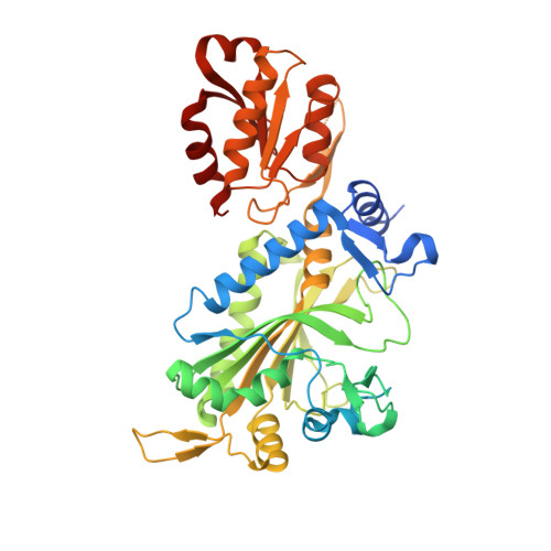 | |
UniProt | |||||
Entity Groups | |||||
| Sequence Clusters | 30% Identity50% Identity70% Identity90% Identity95% Identity100% Identity | ||||
| UniProt Group | G7CIG9 | ||||
Sequence AnnotationsExpand | |||||
Reference Sequence | |||||
| Ligands 5 Unique | |||||
|---|---|---|---|---|---|
| ID | Chains | Name / Formula / InChI Key | 2D Diagram | 3D Interactions | |
| G5A (Subject of Investigation/LOI) Download:Ideal Coordinates CCD File | F [auth A], J [auth B] | 5'-O-(glycylsulfamoyl)adenosine C12 H17 N7 O7 S AMWPZASLDLLQFT-JJNLEZRASA-N |  | ||
| ZN Download:Ideal Coordinates CCD File | H [auth B] | ZINC ION Zn PTFCDOFLOPIGGS-UHFFFAOYSA-N |  | ||
| EDO Download:Ideal Coordinates CCD File | E [auth A] | 1,2-ETHANEDIOL C2 H6 O2 LYCAIKOWRPUZTN-UHFFFAOYSA-N |  | ||
| CL Download:Ideal Coordinates CCD File | D [auth A], I [auth B] | CHLORIDE ION Cl VEXZGXHMUGYJMC-UHFFFAOYSA-M |  | ||
| NA Download:Ideal Coordinates CCD File | C [auth A], G [auth B] | SODIUM ION Na FKNQFGJONOIPTF-UHFFFAOYSA-N |  | ||
| Length ( Å ) | Angle ( ˚ ) |
|---|---|
| a = 171.286 | α = 90 |
| b = 87.388 | β = 104.12 |
| c = 99.152 | γ = 90 |
| Software Name | Purpose |
|---|---|
| PHENIX | refinement |
| Aimless | data scaling |
| XDS | data reduction |
| PHASER | phasing |
| Funding Organization | Location | Grant Number |
|---|---|---|
| National Institutes of Health/National Institute Of Allergy and Infectious Diseases (NIH/NIAID) | United States | 75N93022C00036 |
| National Institutes of Health/Office of the Director | United States | S10OD030394 |


