8U1P | pdb_00008u1p

Local refinement of Plasmodium falciparum gametocyte surface protein Pfs48/45 Domains 1 and 2 in complex with neutralizing antibodies


Experimental Data Snapshot

  • Method: ELECTRON MICROSCOPY
  • Resolution: 3.30 Å
  • Aggregation State: PARTICLE 
  • Reconstruction Method: SINGLE PARTICLE 

Starting Model: experimental
View more details

wwPDB Validation 3D Report Full Report

Validation slider image for 8U1P

This is version 1.2 of the entry. See complete history

Literature

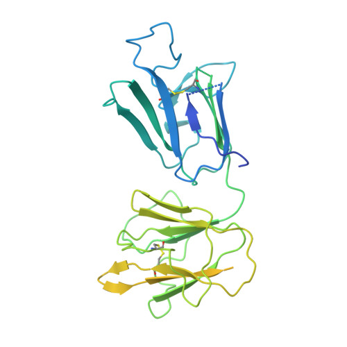
Structural elucidation of full-length Pfs48/45 in complex with potent monoclonal antibodies isolated from a naturally exposed individual.

Kucharska, I.Ivanochko, D.Hailemariam, S.Inklaar, M.R.Kim, H.R.Teelen, K.Stoter, R.van de Vegte-Bolmer, M.van Gemert, G.J.Semesi, A.McLeod, B.Ki, A.Lee, W.K.Rubinstein, J.L.Jore, M.M.Julien, J.P.

(2025) Nat Struct Mol Biol 32: 1396-1407

  • DOI: https://doi.org/10.1038/s41594-025-01532-6
  • Primary Citation Related Structures: 
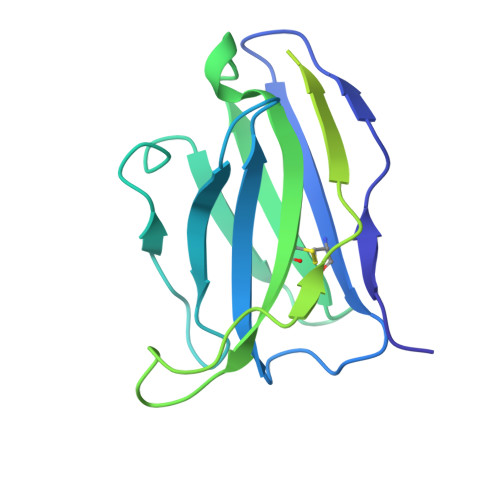
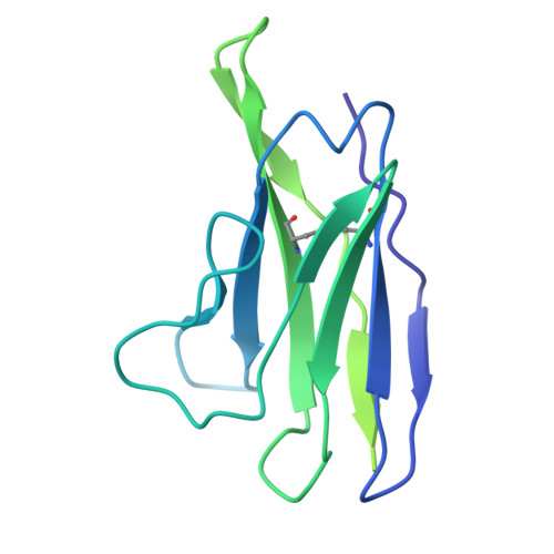
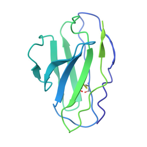
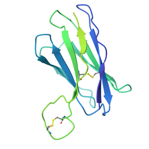
    8U1P, 8U70

  • PubMed Abstract: 

    Biomedical interventions that block the transmission of Plasmodium falciparum (Pf) from humans to mosquitoes may be critical for malaria elimination. Pfs48/45, a gamete-surface protein essential for Pf development in the mosquito midgut, is a target of clinical-stage transmission-blocking vaccines and monoclonal antibodies (mAbs) that disrupt Pf transmission to mosquitoes. Antibodies directed to domain 3 of Pfs48/45 have been structurally and functionally described; however, in-depth information about other inhibitory epitopes on Pfs48/45 is currently limited. Here, we present a cryo-electron microscopy structure of full-length Pfs48/45 in complex with potent human mAbs targeting all three domains. Our data indicate that although Pfs48/45 domains 1 and 2 are rigidly coupled, there is substantial conformational flexibility between domains 2 and 3. Characterization of mAbs against domain 1 revealed the presence of a conformational epitope class that is largely conserved across Pf field isolates and is associated with recognition by potent antibodies. Our study provides insights into epitopes across full-length Pfs48/45 and has implications for the design of next-generation malaria interventions.


  • Organizational Affiliation
    • Program in Molecular Medicine, The Hospital for Sick Children Research Institute, Toronto, Ontario, Canada.

Macromolecule Content 

  • Total Structure Weight: 142.23 kDa 
  • Atom Count: 4,854 
  • Modeled Residue Count: 665 
  • Deposited Residue Count: 1,280 
  • Unique protein chains: 5

Macromolecules

Find similar proteins by:|  3D Structure
Entity ID: 1
MoleculeChains  Sequence LengthOrganismDetailsImage
Gametocyte surface protein P45/48401Plasmodium falciparum 3D7Mutation(s): 0 
Gene Names: PF45/48
UniProt
Find proteins for Q8I6T1 (Plasmodium falciparum (isolate 3D7))
Explore Q8I6T1 
Go to UniProtKB:  Q8I6T1
Entity Groups
Sequence Clusters30% Identity50% Identity70% Identity90% Identity95% Identity100% Identity
UniProt GroupQ8I6T1
Glycosylation
Glycosylation Sites: 3
Sequence Annotations
Expand
Reference Sequence
Find similar proteins by:|  3D Structure
Entity ID: 2
MoleculeChains  Sequence LengthOrganismDetailsImage
RUPA154 Fab Heavy ChainB [auth H]226Homo sapiensMutation(s): 0 
Find similar proteins by:|  3D Structure
Entity ID: 3
MoleculeChains  Sequence LengthOrganismDetailsImage
RUPA154 Fab Light ChainC [auth L]213Homo sapiensMutation(s): 0 
Find similar proteins by:|  3D Structure
Entity ID: 4
MoleculeChains  Sequence LengthOrganismDetailsImage
RUPA58 Fab Light ChainD [auth C]213Homo sapiensMutation(s): 0 
Find similar proteins by:|  3D Structure
Entity ID: 5
MoleculeChains  Sequence LengthOrganismDetailsImage
RUPA58 Fab Heavy ChainE [auth B]227Homo sapiensMutation(s): 0 

Oligosaccharides

Help  
Entity ID: 6
MoleculeChains Length2D Diagram GlycosylationD Interactions
2-acetamido-2-deoxy-beta-D-glucopyranose-(1-4)-2-acetamido-2-deoxy-beta-D-glucopyranoseF [auth D],
G [auth E]
2N-Glycosylation
Glycosylation Resources
GlyTouCan: G42666HT
GlyCosmos: G42666HT
GlyGen: G42666HT

Small Molecules

Ligands 1 Unique
IDChains Name / Formula / InChI Key2D Diagram3D Interactions
NAG

Query on NAG


Download:Ideal Coordinates CCD File
H [auth A]2-acetamido-2-deoxy-beta-D-glucopyranose
C8 H15 N O6
OVRNDRQMDRJTHS-FMDGEEDCSA-N

Experimental Data & Validation

Experimental Data

  • Method: ELECTRON MICROSCOPY
  • Resolution: 3.30 Å
  • Aggregation State: PARTICLE 
  • Reconstruction Method: SINGLE PARTICLE 
EM Software:
TaskSoftware PackageVersion
RECONSTRUCTIONcryoSPARC4
MODEL REFINEMENTPHENIX1.19

Structure Validation

View Full Validation Report


Entry History 

& Funding Information

Deposition Data


Funding OrganizationLocationGrant Number
CIFAR Azrieli Global ScholarsCanada--
Ontario Early Researcher AwardsCanada--
Canada Research ChairsCanada--
Canada Foundation for InnovationCanada--
Ontario Research FundCanada--
Canadian Institutes of Health Research (CIHR)Canada--
Other governmentCanadaCanada Graduate Scholarships Doctoral Award
Netherlands Organisation for Scientific Research (NWO)Netherlands192.061

Revision History  (Full details and data files)

  • Version 1.0: 2025-03-12
    Type: Initial release
  • Version 1.1: 2025-06-04
    Changes: Data collection, Database references
  • Version 1.2: 2025-08-27
    Changes: Data collection, Database references